


A.5. What is regional cardiac tamponade?
Answer:
In contrast to typical cardiac tamponade, where pericardial volume and pressure are evenly distributed around the heart, regional tamponade describes localized cardiac compression due to a loculated intrapericardial collection. This can occur as a result of hematoma formation postcardiotomy or subacutely from effusions developing around adhesions and septation of the pericardial space (Figures 12.2 and 12.3).
Figure 12.2.: Acute Regional Pericardial Effusion: Right Atrial Compression.
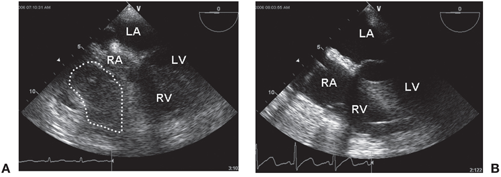
Acute regional pericardial effusion: right atrial compression. The heart is viewed in the midesophageal four-chamber view. A. A loculated pericardial clot (dotted line) is seen around the right atrium (RA), almost obliterating its cavity and partially compressing the right ventricle (RV). B. The same heart following decompression is seen. LA, left atrium; LV, left ventricle.
Figure 12.3.: Acute Regional Pericardial Effusion: Left Atrial Compression.
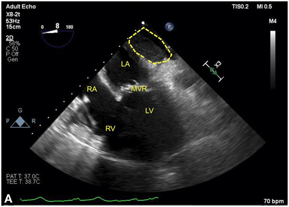
Acute regional pericardial effusion: left atrial compression. A and B show an example of regional pericardial effusion posterior to the heart, compressing the left atrium (LA), is shown. The heart is shown in a midesophageal four-chamber view, with (A) rotated leftward to center the view on the localized pericardial effusion (dotted line). In both views, the left ventricle (LV) is partially obscured by shadowing artifact from a bioprosthetic mitral valve replacement (MVR). A. The LA is partially compressed by the effusion. B. The same heart following decompression, showing restoration of LA size, is seen. Agitated blood can be seen in the right atrium (RA) and right ventricle (RV) during intravenous fluid administration.
The low-pressure RA, right ventricle, and left atrium (LA) are most susceptible to postcardiotomy regional cardiac tamponade. Clot arising from right atrial bleeding typically localizes anteriorly and along the right lateral pericardial recess. Clot can also settle posteriorly behind the LA within the oblique sinus. Following cardiac surgery, the majority of effusions are anterior (especially after coronary artery bypass surgery), whereas posterolateral effusions are more commonly seen following valve replacement. Clinical signs and symptoms in regional cardiac tamponade can be subtle and atypical, corresponding to the discontinuous compression of cardiac chambers. Regional cardiac tamponade has been mistaken for congestive heart failure, acute ventricular dysfunction, septic shock, or pulmonary embolism.
References
- Fowler NO, Gabel M, Buncher CR. Cardiac tamponade: a comparison of right versus left heart compression. J Am Coll Cardiol. 1988;12:187-193.
- Ionescu A, Wilde P, Karsch KR. Localized pericardial tamponade: difficult echocardiographic diagnosis of a rare complication after cardiac surgery. J Am Soc Echocardiogr. 2001;14:1220-1223.
- Russo AM, O'Connor WH, Waxman HL. Atypical presentations and echocardiographic findings in patients with cardiac tamponade occurring early and late after cardiac surgery. Chest. 1993;104:71-78.