section name header

Basic Information

AUTHORS: Omar Karim, BS and Caroline P. Meehan, MD

Definition

Gout refers to a group of disease states caused by deposition of monosodium urate (MSU) in tissue, resulting from prolonged hyperuricemia. Clinical manifestations of gout include acute and chronic arthritis, soft tissue inflammation, tophus formation, gouty nephropathy, and nephrolithiasis. Untreated hyperuricemia in patients with gout may lead to chronic destructive deforming arthritis.

ICD-10CM CODES
M10Gout
M10.0Idiopathic gout
M10.1Lead-induced gout
M10.2Drug-induced gout
M10.3Gout due to impairment of renal function
M10.4Other secondary gout
M10.9Gout, unspecified
Epidemiology & Demographics
Prevalence

Self-reported prevalence in the U.S. is estimated at 3.9% of adults.1 In the U.S., gout has been diagnosed in more than 10 million adults.2

Predominant Sex

Male:female ratio 4:1. However the prevalence of gout among women has increased in past decade. Compared with men, gout in women is more likely to be associated with diabetes, coronary heart disease, and chronic kidney disease.3

Predominant Age

30 to 50 yr in men; >60 yr in women

Etiology

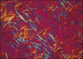
  • Gout is caused by inflammation resulting from MSU crystal deposition. The primary risk factors for MSU deposition are hyperuricemia, although local factors such as temperature, pH, and mechanical stress may play a role. Figs. E1, E2, and E3 illustrate the pathophysiology of gout.
  • Hyperuricemia and gout develop from excessive uric acid production, a decrease in the renal excretion of uric acid, or both.
  • Primary hyperuricemia results from an inborn error of metabolism and may be attributed to several biochemical defects.
  • Secondary hyperuricemia may develop as a complication of acquired disorders (e.g., leukemia) or as a result of the use of certain drugs (e.g., diuretics). Consumption of alcohol, especially beer, increases the risk of gout, and fructose-rich beverage intake is associated with hyperuricemia. Gout promoters and inhibitors are summarized in Table 1.

TABLE 1 Gout Promoters and Inhibitors

Crystal formationSeed nucleus (particulate)
Immunoglobulin
Phagocytes
Low temperature
Low pH
Cation concentration
Intraarticular dehydration
Other (unknown) macromolecules
Triggering the acute flare (local factors)Rapid change in urate level
Microcrystal release
IgG coat (apolipoproteins B, E inhibitory)
Complement activation (classical, alternate, MAC)
Inflammasome activation
Cytokine and chemokine release
Endothelial activation (e-selectin, ICAM-1, VCAM-1)
Local trauma
Presence of susceptible phagocytes, mast cells (systemic events)Surgery, trauma
Infections, other intercurrent systemic illness
Alcohol, dietary intake
Drugs that raise or lower circulating urate level

ICAM-1, Intercellular adhesion molecule 1; Ig, immunoglobulin; MAC, membrane attack complex; VCAM-1, vascular cell adhesion molecule 1.

A diverse array of proteins and other mediators have been identified on the surfaces of urate crystals. In addition to their proinflammatory effect through opsonizing existing crystals, immunoglobulin M (IgM) and immunoglobulin G (IgG) antibodies may promote crystal formation by providing a stable molecular platform for crystal nucleation and growth. Apolipoproteins are antiinflammatory molecules that coat crystals. The characteristics of the phagocytes encountering the crystals may be crucial; macrophages that are more differentiated are less likely to elicit proinflammatory cytokines.

From Hochberg MC et al: Rheumatology, ed 7, Philadelphia, 2019, Elsevier.

Figure E1 Many signals impact the critical points in this pathway.

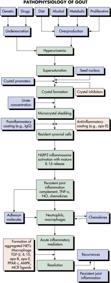
Patients with hyperuricemia do not necessarily form urate crystals. Crystals ingested by phagocytes sometimes elicit remarkably little inflammation, and the inflammation is often low grade or clinically silent in advanced gout, indicating robust regulation of the pathways shown. Factors leading to the spontaneous termination of the acute gout flare are incompletely understood. AMPK, Adenosine monophosphate-activated protein kinase; apo, apolipoprotein; IgG, immunoglobulin G; IL, interleukin; MCR, melanocortin receptor; NET, neutrophil extracellular trap; NLRP3, nucleotide-binding oligomerization domain-like receptor 3; NO, nitric oxide; PPAR, peroxisome proliferator-activated receptor; TGF-β, transforming growth factor-β; TNF-α, tumor necrosis factor-α.

From Hochberg MC: Rheumatology, ed 7, Philadelphia, 2019, Elsevier.

Figure E2 Initial phases of monosodium urate (MSU) crystal-induced activation.

IL, Interleukin.

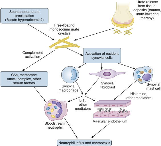
The presence of “fresh” MSU crystals, resulting either from spontaneous precipitation or the liberation of crystals from established pools, results in direct complement activation and activation of resident cells in the synovium including macrophages, fibroblasts, and mast cells. Activated cells produce IL-1β and other cytokines, as well as multiple other mediators (not all illustrated) that in turn activate both bloodstream neutrophils and endothelial cells. These responses permit neutrophils to adhere to and traverse the endothelium, resulting in neutrophil influx and the further propagation of inflammation as neutrophils undergo direct activation by MSU crystals (not illustrated). See text for additional details.

From Firestein GS et al: Firestein & Kelley’s textbook of rheumatology, ed 11, Philadelphia 2021, Elsevier.

Figure E3 Propagation of the acute gouty response by activated neutrophils.

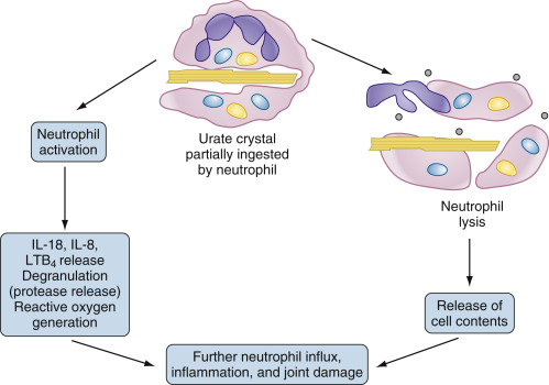
Neutrophils that enter the joint migrate toward and phagocytose crystals. In the case of crystals coated with immunoglobulins and complement, the resultant activation results in synthesis and/or release of inflammatory mediators such as interleukin-1β (IL-1β), IL-8, and tumor necrosis factor, as well as proteases and reactive oxygen species. In the case of uncoated crystals, the crystal frequently interacts with and lyses the membrane of the phagolysosome, spilling toxic contents and leading to cell lysis. In both cases, the result is local tissue damage and recruitment of additional neutrophils from the bloodstream in an explosive inflammatory cycle. LTB4, Leukotriene B4.

From Firestein GS et al: Firestein & Kelley’s textbook of rheumatology, ed 11, Philadelphia 2021, Elsevier.

Physical Findings & Clinical Presentation
Acute Gout

  • Rapid onset of pain and swelling and erythema of a distal joint and/or periarticular soft tissue. Box 1 summarizes clinical pearls in acute gout attacks.
  • May present as monoarthritis of any joint. Acute gout of the first metatarsophalangeal (MTP) joint is known as podagra.
  • 10% to 15% of attacks are polyarticular.
  • Spontaneous resolution occurs over days to weeks.

BOX 1 Acute Gout: Clinical Pearls

  • Abrupt and rapid onset
  • Maximal symptom intensity at 8-12 h
  • Attacks often come on at night or early morning
  • Most commonly monoarticular in men during first attack
  • Metatarsophalangeal joint is first affected in 50% of cases; other commonly affected joints: Ankle, heel, knee, wrists, and hands
  • Joints are red, hot, swollen, and exquisitely tender
  • Attacks resolve within days to weeks without treatment

From Hochberg MC et al: Rheumatology, ed 7, Philadelphia, 2019, Elsevier.

Chronic Tophaceous Gout (Fig. E1)

  • Insidious onset of painless arthritis and soft tissue swelling
  • Distal small joints characteristic
  • May be confused with nodal osteoarthritis
  • Box 2 summarizes clinical pearls in chronic gout

BOX 2 Chronic Gout: Clinical Pearls

  • Pattern of symptom changes: Time between attacks shortens; more joints may be involved
  • Tophaceous disease may result in a destructive arthropathy
  • Tophaceous deposits may be bothersome and cause marked reduction in quality of life
  • Increased risk for nephrolithiasis
  • Associated with comorbidities such as metabolic syndrome, diabetes mellitus, chronic kidney disease, cardiovascular disease, hyperlipidemia, and obesity

From Hochberg MC et al: Rheumatology, ed 7, Philadelphia, 2019, Elsevier.

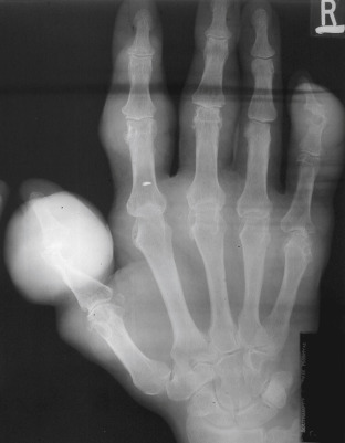
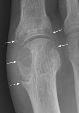
Figure E4 Advanced tophaceous gout with multiple intra-articular and periarticular tophi.

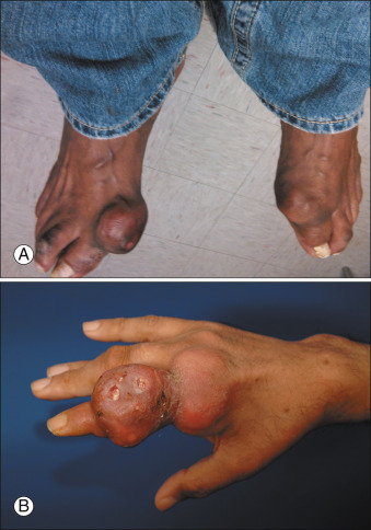
Tophi observed on bilateral first metatarsophalangeal joints (A) (right greater than left) and the hand (B) in a patient with severe tophaceous gout.

From Hochberg MC et al: Rheumatology, ed 7, Philadelphia, 2019, Elsevier.

Diagnosis

Differential Diagnosis of Acute Gout

  • Septic arthritis, cellulitis
  • Pseudogout, calcium pyrophosphate crystal deposition disease
  • Trauma
Differential Diagnosis of Chronic Gout

  • Osteoarthritis (OA; especially nodal OA in women)
  • Rheumatoid arthritis
  • Psoriatic arthritis

Section II describes the differential diagnosis of acute monoarticular and oligoarticular arthritis.

Workup

Arthrocentesis and examination of synovial fluid

Laboratory Tests

  • Uric acid: All patients with gout are hyperuricemic at some time, but during an acute attack the serum uric acid may be normal or low.
  • Synovial aspirate: Usually cloudy and markedly inflammatory in nature (elevated white blood cells). Urate crystals in fluid are needle shaped and strongly negatively birefringent under polarized microscopy 4 (Fig. E5).
  • CBC: Neutrophilic leukocytosis often present.
  • Inflammatory markers: Erythrocyte sedimentation rate and C-reactive protein often elevated.

Figure E5 Monosodium urate crystals from tophus.

Crystals aspirated from a subcutaneous tophus. Polarized transmission white (halogen) light; 90-degree extinction filter; 530-nm first-order compensator.

From Hochberg MC et al: Rheumatology, ed 7, Philadelphia, 2019, Elsevier.

Imaging Studies

  • Plain radiography for diagnosis and evaluation. No typical findings in early gouty arthritis, but late disease is associated with characteristic punched-out marginal erosions (Fig. E6) and overhanging edges.
  • Musculoskeletal ultrasound has been shown to be an effective means of detecting monosodium urate crystal deposition. Ultrasound can differentiate urate crystals that are found on the surface of articular cartilage from CPPD crystals that are seen within the substance of the cartilage (Fig. E7). Double contour sign can be seen.5
  • Dual-energy computed tomography (DECT) allows for color-coded images and detection of gout deposits and may be beneficial if there is difficulty in establishing the diagnosis.

Figure E6 Chronic tophaceous gout. Asymmetric large eccentric soft tissue lumps (tophi).

Underlying gouty erosions are seen at several sites, particularly the distal interphalangeal joint of the right little finger and the carpometacarpal joint of the thumb.

From Adam A et al: Grainger & Allison’s diagnostic radiology, ed 5, London, 2007, Churchill Livingstone; in Grant LA: Grainger & Allison’s diagnostic radiology essentials, ed 2, Philadelphia 2019, Elsevier.

Figure E7 Classic radiographic findings of gout in a 69-yr-old man.

Note the asymmetric, well-marginated erosions in the head of the first metatarsal and lesser erosions in the opposing phalanx (solid arrows). The medial erosions are characteristically larger than the lateral erosions. There is a “hook” sign on the inferior margin of the large medial erosion of the head of the metatarsal (dashed arrow). The joint space is preserved, and there is no osteopenia.

From Pope TL et al: Musculoskeletal imaging, ed 2, Philadelphia, 2014, WB Saunders.

Treatment

Treatment Options for Acute Gout (Table 2

  • Nonsteroidal antiinflammatory medication (Table 3).
    1. Indomethacin 75 mg bid
    2. Ibuprofen 800 mg tid
    3. Naproxen 500 mg bid
    4. Celecoxib 200 mg bid
  • Low-dose colchicine (less toxic, as effective as traditional high-dose colchicine): 1.2 mg PO, followed by 0.6 mg PO 1 h later, then 0.6 mg/day or bid. Dose for renal function.6
  • Intraarticular corticosteroid injection (treatment of choice for monoarticular large joint attack): Triamcinolone acetonide 40 mg or equivalent for knee.
  • Systemic corticosteroid therapy: Prednisone 40 mg PO for 3 days, then taper over 10 days (limited evidence currently, but studies have shown efficacy).7

TABLE 2 Therapeutic Options for Acute Gouty Inflammation

Drug OptionLevel of EvidenceTypical Regimens
COX-nonselective NSAIDsGrade A evidence for each regimen listedNaproxen, 750-1000 mg/day PO in divided doses for 3 days; then 500-750 mg total daily for 4-7 days
Sulindac, 300-400 mg/day in divided doses for 7-10 days
Indomethacin, 150-200 mg/day PO in divided doses for 3 days; then 100 mg/day PO in divided doses for 4-7 days
COX-2-selective NSAIDsGrade AFor example: Celecoxib, 800 mg; then 400 mg on day 1, followed by 400 mg bid for 7 days
Systemic corticosteroidsGrade A evidence for oral prednisolone, 35 mg/day for five or six doses; lower evidence grades for other regimensPrednisone, 30-60 mg/day for 3 days; then taper every 3 days by 10-15 mg less as a daily dose until discontinuation
Oral prednisolone, 35 mg/day for 5-7 days
Medrol dose pack (for less severe flares) or to initiate therapy
In an NPO patient:
  • Triamcinolone, 60 mg IM once with additional corticosteroid treatment as needed
  • Methylprednisolone, 100-150 mg IV for 1-2 days
Intraarticular corticosteroidsGrade C evidenceParticularly useful for flares in a single large joint
Triamcinolone acetonide preparations are especially useful
Dose titrated to the size of the joint
ACTHGrade C evidence25 USP units of synthetic ACTH SC for less severe flares; 40 USP units IM or IV once for more severe flares (including larger joint flares and polyarticular gout)
One or two repeated doses of synthetic ACTH at intervals of 12 h are often required with each of these regimens
Oral colchicineGrade A evidence for FDA-approved regimenIn the U.S: To treat an early acute gout flare: 1.2 mg once followed by 0.6 mg in 1 h and then 12 h later; oral low-dose colchicine at prophylaxis doses until the acute gout flare resolves
Outside the U.S: 0.5 mg tid for several days is the EULAR-recommended dosing regimen
Oral colchicine treatment of acute gout should be limited to once every 2 wk in those already taking a maintenance low dose of colchicine
Off-label use in U.S: IL-1 antagonismGrade C for anakinra; grade A for canakinumabUse of anakinra (e.g., 100 mg/day SC for 3 days) or canakinumab (150 mg SC as single dose) for acute gout flare is not approved by the FDA, but canakinumab is approved by the EMA for this indication

ACTH, Adrenocorticotropic hormone; bid, twice a day; COX, cyclooxygenase; EMA, European Medicines Agency; EULAR, European League Against Rheumatism; FDA, U.S. Food and Drug Administration; IL-1, interleukin-1; IM, intramuscularly; IV, intravenously; NPO, nothing by mouth; NSAID, nonsteroidal antiinflammatory drug; PO, by mouth; SC, subcutaneously; tid, three times a day; USP, U.S. Pharmacopeia.

From Hochberg MC et al: Rheumatology, ed 7, Philadelphia, 2019, Elsevier.

TABLE 3 Treatment of Gout

Acute GoutInterval GoutTreatment of Hyperuricemia
NSAIDs (preferred): Indomethacin 50 mg qid or ibuprofen 800 mg tid (or other NSAID in full doses). Contraindicated in patients with renal insufficiency and gastrointestinal disorders.
Or
Colchicine, oral: 1.2 mg followed by a second dose of 0.6 mg 1 h later.
Contraindicated in patients with renal insufficiency and gastrointestinal disorders
Or
Intraarticular steroids (Treatment of choice for large joint monoarthritis):
Triamcinolone 40 mg or equivalent for knee
Or
Systemic steroid therapy (for patients in whom NSAIDs and colchicine are contraindicated)
Prednisone 30-50 mg/day PO or in divided doses. May use lower dose in diabetic or postsurgical patients.
Colchicine, oral: 0.6-1.2 mg/day as prophylaxis against recurrent attacks.
NSAIDs may also be used for prophylaxis.
Hypouricemic agent: Indicated for patients with recurrent attacks despite prophylaxis, severe hyperuricemia, presence of tophi, urolithiasis, or gouty arthritis
Other: Weight loss, reduce alcohol (especially beer), diet low in seafood, red meat, organ meat, and fructose
Colchicine, oral: 0.6-1.2 mg/day for 4-6 wk before initiating hypouricemic therapy and for several months afterward to prevent recurrent attacks during initiation of hypouricemic therapy
And
Allopurinol: Initial dose 100 mg/day in patients with renal insufficiency or very high uric acid levels. Increase dose as needed to attain uric acid less than 6 mg/dl
Or
Uricosuric agent (Use only in patients with good renal function and <600 mg uric acid in a 24-h collection): Probenecid, 0.5-1 g bid, or sulfinpyrazone 100 mg tid or qid
Other: Consider febuxostat for patients allergic to allopurinol and the addition of lesinurad in patients resistant to xanthine oxidase inhibitors. Pegloticase may be useful for selected patients with severe tophaceous gout

bid, Twice a day; NSAID, nonsteroidal antiinflammatory drug; PO, by mouth; qid, four times a day; tid, three times a day.

Treatment of Hyperuricemia in Patients With Gout

The American College of Rheumatology, as well as most international rheumatology guidelines, recommend that every patient with gout who has tophi, more than two attacks of gout per year, chronic kidney disease, or nephrolithiasis be treated with pharmacologic urate-lowering therapy. Serum uric acid should be monitored on a regular basis and urate-lowering therapy intensified until a target of less than 6 mg/dl is reached. In most cases, urate-lowering therapy should be continued for life. Fig. E8 summarizes pharmacologic serum urate-lowering treatment.1

The American College of Physicians Guidelines recommends a more conservative approach based on recurrence of symptoms. These guidelines have been criticized for ignoring the progressive nature of gout and perpetuating the well-documented underuse and under-dosing of urate-lowering therapy.

Figure E8 Algorithm for Pharmacologic Serum Urate-Lowering Treatment of Hyperuricemia in Patients with a Confirmed Diagnosis of Gout

The algorithm, discussed in the text, summarizes the first-, second-, and third-line approaches to pharmacologic urate-lowering therapy, including management of refractory hyperuricemia in difficult gout. CKD, Chronic kidney disease; PCR, polymerase chain reaction.

From Hochberg MC et al: Rheumatology, ed 7, Philadelphia, 2019, Elsevier.

Nonpharmacologic Therapy

Lifestyle and dietary modification should always be a component of therapy for patients with gout, but this is rarely effective without concomitant pharmacologic urate-lowering therapy, as dietary modification can lower uric acid only about 1 mg/dl. Recommendations include reducing ingestion of red meat, kidney, liver, yeast extract, shellfish, and overall protein along with restricting alcohol intake. Discontinuation of diuretic therapy may help lower serum uric acid. Women with normal weight and best adherence to dietary approaches to stop hypertension (DASH) diet have a 68% lower risk for gout than women who are overweight or obese and are least adherent to the DASH diet.3

Pharmacologic Treatment of Symptomatic Hyperuricemia
Allopurinol

Allopurinol is very effective and safe when used properly. Correct dosing and patient compliance are essential elements in the prevention of erosive and tophaceous gout. Patients with renal insufficiency are at increased risk for allopurinol hypersensitivity, which manifests as fever, rash, and hepatitis occurring most commonly in the first 3 months of therapy. The rash may progress to life-threatening toxic epidermal necrolysis if not recognized early.

Traditionally, therapy with allopurinol is initiated several weeks after the acute attack has resolved. However, initiation of allopurinol at presentation may improve long-term compliance without reducing the efficacy of acute treatment. The initial dose should be low (100 mg/day depending on creatinine clearance) in patients with renal insufficiency and those with very high uric acid levels.8 High initial doses are associated with increased incidence of allopurinol hypersensitivity. The serum uric acid should be reevaluated after 4 to 6 wk of therapy, and the allopurinol dose adjusted to reduce the serum uric acid to <6 mg/dl. The most common therapeutic dosage of allopurinol is 300 mg/day, but the dose may be increased by 50 to 100 mg every 2 to 3 wk until the target serum uric acid level is achieved. There is evidence that increasing allopurinol doses in patients with renal insufficiency does not result in significant toxicity, but concurrent use with statins and colchicine is associated with a higher incidence of adverse effects. Some authors have reported using doses as high as 800 mg/day without excess toxicity. It is recommended that patients of Han Chinese, Thai, and Korean ancestry be tested for HLA-B5801 before initiating allopurinol as these individuals are at high risk of allopurinol hypersensitivity if this allele is present.1

Febuxostat

Febuxostat is a xanthine oxidase inhibitor that has been shown to be more potent than allopurinol 300 mg/day for reducing serum uric acid. The chemical structure of febuxostat is different from allopurinol, making cross-reactive allergy unlikely. The metabolism of febuxostat is primarily hepatic, which obviates the need for dose adjustments because of renal insufficiency. Some cases of hepatic toxicity have been reported, and it is recommended that liver function tests be monitored. Febuxostat may help preserve renal function in patients with chronic kidney disease (CKD) but has not been tested in patients with severe renal failure.1

The primary indication for febuxostat is demonstrated allergy to allopurinol. The cost of febuxostat may be as much as 40 times that of allopurinol, and there is some evidence suggesting that febuxostat may be associated with higher cardiovascular and all-cause mortality than allopurinol in patients with cardiovascular risk factors. In 2019 the FDA added a boxed warning for increased risk of death with febuxostat.9

Probenecid

Uricosuric agents may be used in patients with good renal function and urinary uric acid <600 mg in a 24-h collection. Probenecid can be used in patients with intolerance to xanthene-oxidase inhibitors. Compliance is poor due to the necessity of taking the drug more often than once daily.1

Lesinurad

Lesinurad is a URAT1 and OAT4 inhibitor approved by the FDA in 2015 for gout-associated hyperuricemia unresponsive to xanthine oxidase inhibitor monotherapy, to be taken in combination with a xanthine oxidase inhibitor.10 This drug was withdrawn in 2019 because of business-related reasons without safety concerns noted.

Pegloticase

Intravenous PEGylated uricase is FDA approved for treatment of severe refractory tophaceous gout. It is a PEGylated recombinant mammalian uricase that rapidly degrades urate when given intravenously. Use is limited by very high cost and potential toxicities, including frequent gout flares and anaphylaxis. Rasburicase is a nonpegylated uricase used in tumor lysis syndrome and is not indicated for gout treatment.11

Referral

  • Rheumatologist if diagnosis is not clear or therapy is complicated
  • Podiatrist for management of pedal complications

Pearls & Considerations

Patient & Family Education

It is essential that patients, families, physicians, and other members of the health care team appreciate the importance of compliance with a daily allopurinol regimen if recurrent flares and progression to chronic arthritis and tophi are to be avoided. Allopurinol should be discontinued only for symptoms suggesting hypersensitivity; otherwise, it should be continued during flares, medical illnesses, and surgical procedures.14

Related Content

Gout (Patient Information)

Hyperuricemia (Related Key Topic)

Related Content

    1. Khanna D. : American College of Rheumatology guidelines for management of gout. Part 1: systematic non-pharmacologic and pharmacologic therapeutic approaches to hyperuricemiaArthritis Care Res. ;64(10):1431-1446, 2012.
    2. Mikuls T.R. : GoutN Engl J Med. ;387(20):1877-1887, 2022.
    3. Yokose C. : Adherence to 2020 to 2025 dietary guidelines for Americans and the risk of new-onset female goutJAMA Intern Med. ;182(3):254-264, 2022.
    4. Newberry S.J. : Diagnosis of gout: a systematic review in support of an American College of Physicians clinical practice guidelineAnn Intern Med. ;166:27-36, 2017.
    5. Ogdie A. : Performance of ultrasound in the diagnosis of gout in a multicenter study: comparison with monosodium urate monohydrate crystal analysis as the gold standardArthritis Rheumatol. ;69:429-438, 2017.
    6. Terkeltaub R.A. : High versus low dosing of oral colchicine for early acute gout flareArthritis Rheum. ;62:1060-1068, 2010.
    7. Hudson T. : Oral prednisone in the treatment of goutAnn Intern Med. ;164:464-471, 2016.
    8. Bathini L. : Initiation dose of alopurinol and the risk of severe cutaneous reactions in older adults with CKD: a population-based cohort studyAm J Kidney Dis. ;80(6):730-739, 2022.
    9. White W.B. : Cardiovascular safety of febuxostat or allopurinol in patients with goutN Engl J Med. ;378(13):1200-1210, 2018.
    10. Dalbeth N. : Lesinurad, a selective uric acid reabsorption inhibitor, in combination with febuxostat in patients with tophaceous gout: findings of a phase III clinical trialArthritis Rheumatol. ;69(9):1903-1913, 2017.
    11. Sundy J.S. : Efficacy and tolerability of pegloticase for the treatment of chronic gout in patients’ refractory to conventional treatment: two randomized controlled trialsJ Am Med Assoc. ;306:711-720, 2011.
    12. Yokose C. : Adherence to 2020 to 2025 dietary guidelines for Americans and the risk of new-onset female goutJAMA Intern Med. ;182(3):254-264, 2022.
    13. Liew J.W. : Use of anakinra in hospitalized patients with crystal-associated arthritisJ Rheumatol. ;46(10):1345-1349, 2019.
    14. Lieke E.J.M. : Medication adherence among gout patients initiated allopurinol: a retrospective cohort study in the Clinical Practice Research Datalink (CPRD)Rheumatology. ;57:1641-1650, 2018.