Author: Fred F. Ferri, MD


Cholecystitis is acute or chronic inflammation of the gallbladder generally caused by gallstones (>95% of cases).
| ICD-10CM CODES | |||
| K81.9 | Acute cholecystitis | ||
| K80.00 | Calculus of gallbladder with acute cholecystitis without obstruction | ||
| K81.9 | Cholecystitis, unspecified | ||
- •Acute cholecystitis occurs most commonly in women during the fifth and sixth decades. Approximately 120,000 cholecystectomies are performed for acute cholecystitis annually in the U.S.
- •The incidence of gallstones is 0.6% in the general population and much higher in certain ethnic groups (>75% of Native Americans by age 60 yr). Most patients with gallstones are asymptomatic. Of such patients, biliary colic develops in 1% to 4% annually.
- •Pain and tenderness in the right hypochondrium or epigastrium; pain possibly radiating to the infrascapular region
- •Palpation of the right upper quadrant eliciting marked tenderness and stoppage of inspired breath (Murphy sign)
- •Guarding
- •Fever (33%)
- •Jaundice (25% to 50% of patients)
- •Palpable gallbladder (20% of cases)
- •Nausea and vomiting (>70% of patients)
- •Fever and chills (>25% of patients)
- •Medical history often revealing ingestion of large, fatty meals before onset of pain in the epigastrium and right upper quadrant
- •Gallstones (>95% of cases). Factors contributing to gallstone formation are illustrated in Fig. E1
- •Ischemic damage to the gallbladder, critically ill patient (acalculous cholecystitis)
- •Infectious agents, especially in patients with AIDS (cytomegalovirus, Cryptosporidium)
- •Strictures of the bile duct
- •Neoplasms, primary or metastatic
- •Risk factors for cholelithiasis include age, obesity, female sex, rapid weight loss, ethnicity/race (Native American), use of contraceptives, pregnancy, diabetes mellitus, use of glucagon-like peptide-1 (GLP-1) receptor agonists,1 hemolysis, total parenteral nutrition, biliary parasites
Figure E1 Five Primary Defects Work Together to Promote Formation of Cholesterol Gallstones
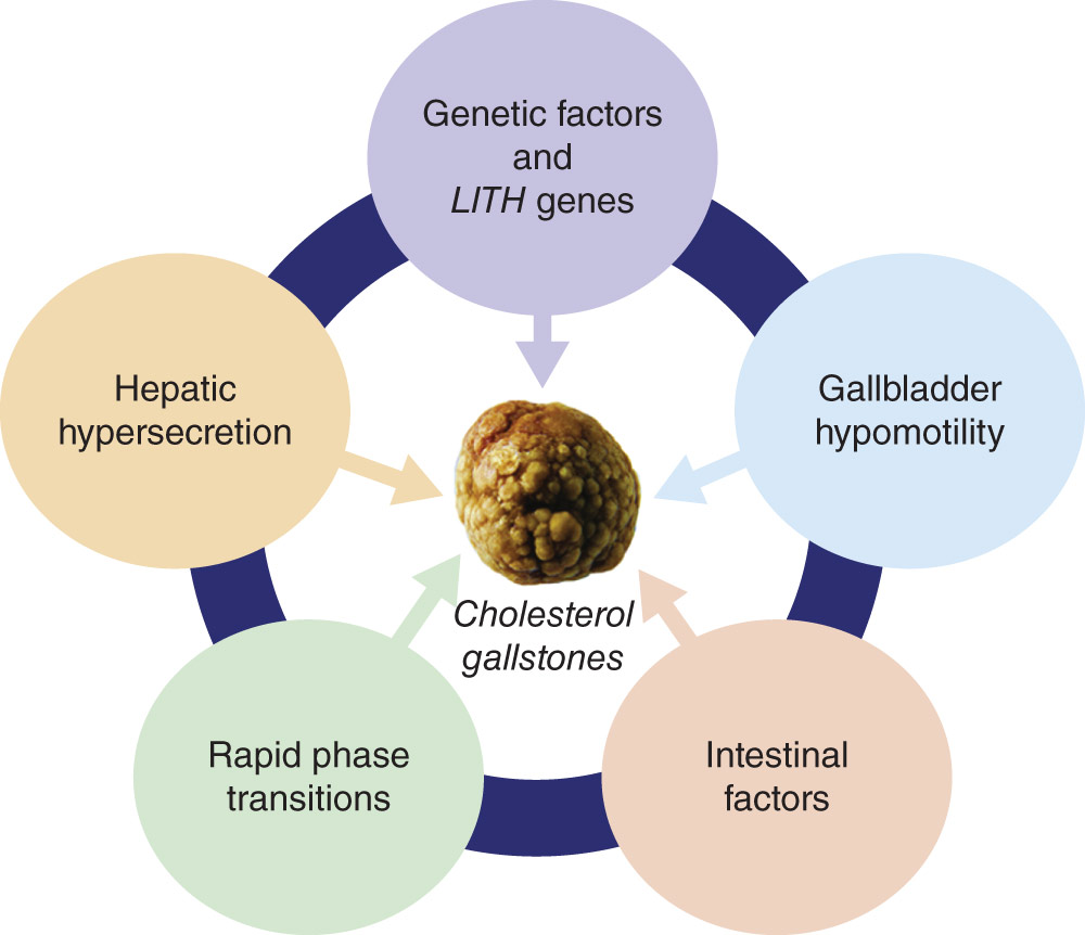
The five defects are genetic factors and LITH (gallstone) genes, hepatic hypersecretion of cholesterol, gallbladder hypomotility, rapid phase transitions, and intestinal factors. The hypothesis proposed is that hepatic hypersecretion of biliary cholesterol is the primary defect and is the outcome, in part, of a complex genetic predisposition. Downstream effects include gallbladder hypomotility and rapid phase transitions. A major result of gallbladder hypomotility is alteration in the kinetics of the enterohepatic circulation of bile salts (intestinal factors). Alterations in intestinal factors result in increased cholesterol absorption, as well as reduced bile salt absorption, that leads to abnormal enterohepatic circulation of bile salts and a diminished biliary bile salt pool size. Not only does gallbladder hypomotility facilitate cholesterol nucleation and crystallization, but it also allows the gallbladder to retain solid platelike cholesterol monohydrate crystals. Although a large number of candidate LITH genes have been identified in mouse models and many human LITH genes have been discovered, their contributions to gallstone pathogenesis require further investigation.
(From Feldman M et al: Sleisenger and Fordtran’s gastrointestinal and liver disease, ed 11, Philadelphia, 2021, Elsevier.)