section name header

Basic Information

Author: Fred F. Ferri, MD

Definition

Choledocholithiasis is a derivation from the Greek words of choli (bile), docheion (container), and lithos (stone) and refers to the presence of gallstones within the common bile duct (CBD).

Synonym

  • Common bile duct stone(s)
ICD-10CM CODE
K80.50Calculus of bile duct
Epidemiology & Demographics

  • While the exact incidence and prevalence are unknown, an estimated 10% to 20% of patients are found to have choledocholithiasis at the time of cholecystectomy.
  • Passage of gallstones into the CBD occurs in approximately 10% to 15% of those with cholelithiasis, and the incidence is known to increase with age. All factors that promote migration of secondary gallstones into the CBD are not well understood; however, a cystic duct diameter greater than 4 mm is associated with higher risk of migration. Approximately 95% of those with choledocholithiasis will also have cholelithiasis.
  • Risk factors for gallstone formation include nonmodifiable factors such as age, female sex, family history, ethnic background, and genetic predilection, while modifiable factors are centripetal obesity and metabolic syndrome, rapid weight loss, ileal Crohn disease, cirrhosis, total parenteral nutrition, and medications such as estrogen replacement therapy.
Physical Findings & Clinical Presentation

  • Uncomplicated choledocholithiasis presents with biliary colic; it is classically described as intense and constant pain in the right upper quadrant or epigastric region, associated with nausea and vomiting.
  • Occasionally patients may remain asymptomatic, but resolution of pain more often reflects passage of stone into the bowel.
  • Physical examination demonstrates right upper quadrant or epigastric tenderness and occasionally jaundice.
  • Courvoisier sign for a palpable gallbladder is more typically associated with malignant obstruction of the CBD, but it has been reported with choledocholithiasis.
  • Other clinical findings of fever (Charcot triad), hypotension, and altered mental status (Reynolds pentad) are found only when choledocholithiasis is complicated by acute cholangitis.
  • Choledocholithiasis can also be complicated by acute pancreatitis.
Etiology

  • The majority of cases are due to passage of cholesterol stones from the gallbladder into the common bile duct.
  • De novo formation of choledocholithiasis (primary choledocholithiasis) is uncommon but is seen among those with increased propensity for pigment stone formation due to chronic recurrent pyogenic cholangitis from trematodes, congenital biliary duct anomalies such as peripapillary diverticulum or abnormal ductal angulation, dilated or strictured ducts or MDR3 gene defects causing impairments in biliary phospholipid secretions, or biliary stasis such as from cystic fibrosis.
  • Brown pigment stones comprise the majority of pigment stones in the bile duct, often found proximal to biliary stricture and associated with cholangitis.

Diagnosis

Differential Diagnosis

  • Biliary pain
  • Acute cholecystitis
  • Sphincter of Oddi dysfunction
  • Functional gallbladder disorder
  • Malignant obstruction
  • Choledochal cyst
  • Papillary stenosis
  • AIDS-associated cholangiopathy

Choledocholithiasis can easily be differentiated from these other etiologies based on thorough history, physical examination, and imaging studies.

Workup

  • The constellation of symptomatic cholelithiasis with elevated liver enzymes should prompt a transabdominal ultrasound (US) of the right upper quadrant to evaluate for a stone in the CBD, which is the most reliable predictor of choledocholithiasis.
  • Elevated liver enzymes are 94% sensitive in detecting choledocholithiasis.
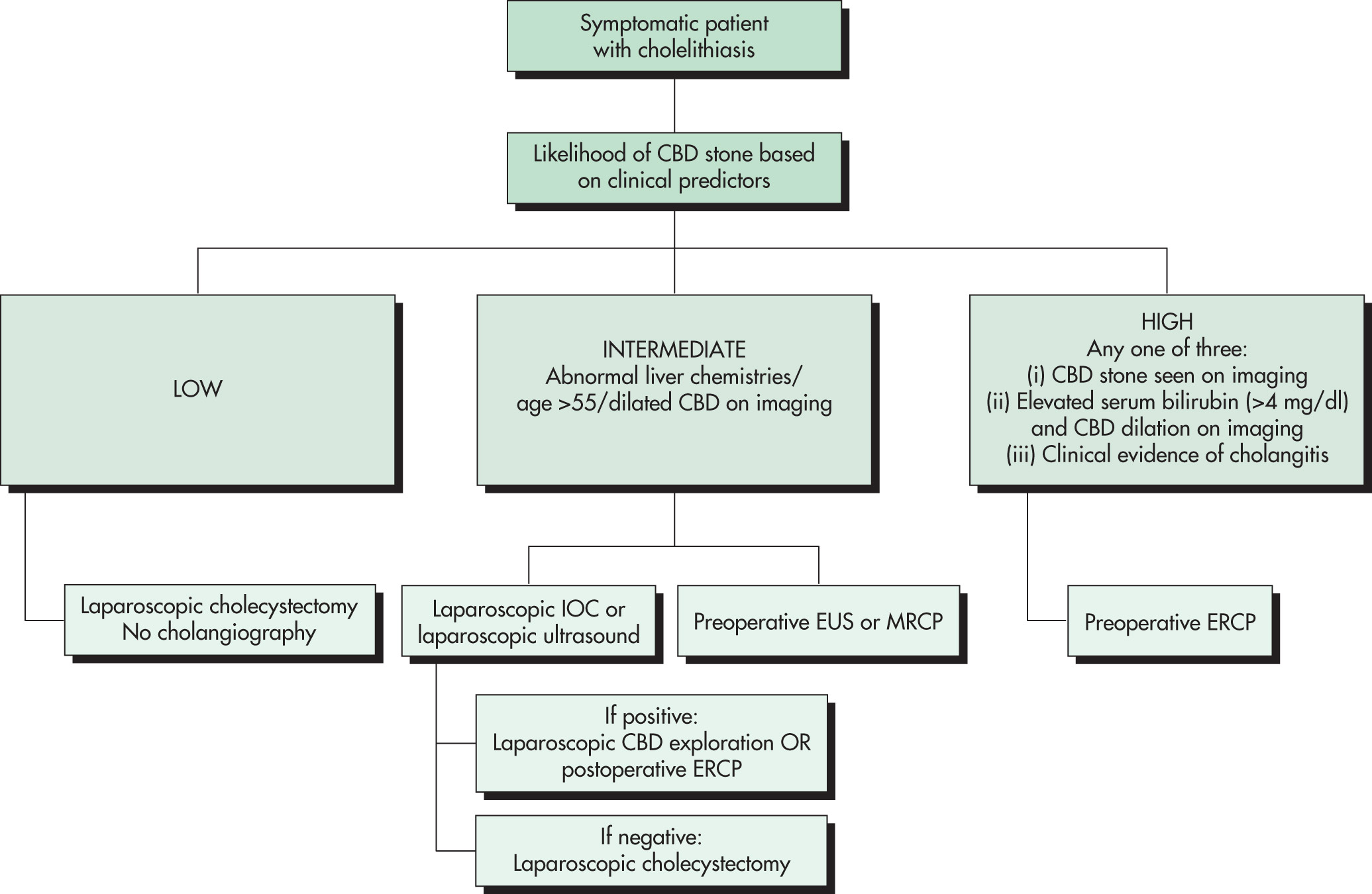
  • Clinical predictors should be utilized to risk-stratify patients and to inform the next step in management.
  • High-risk predictors of choledocholithiasis:
    1. 1.CBD stone seen on US or cross-sectional imaging
    2. 2.Elevated serum bilirubin (>4 mg/dl) with evidence of dilated CBD on imaging
    3. 3.Clinical or biochemical evidence of ascending cholangitis
  • Intermediate-risk predictors of choledocholithiasis include abnormal liver biochemistries, age >55 yr, and/or a dilated CBD seen on imaging.
  • See Fig. E1 for a proposed risk stratification model based on American Society for Gastrointestinal Endoscopy guidelines.

Figure E1 Algorithm for the management of patients with symptomatic cholelithiasis.

!!flowchart!!

CBD, Common bile duct; ERCP, endoscopic retrograde cholangiopancreatography; EUS, endoscopic ultrasound; IOC, intraoperative cholangiogram; MRCP, magnetic resonance cholangiopancreatography.

(Adapted from Feldman M et al: Sleisenger and Fordtran’s gastrointestinal and liver disease, ed 11, Philadelphia, 2021, Elsevier; and Buxbaum J et al: ASGE guideline on the role of endoscopy in the evaluation and management of choledocholithiasis, Gastrointest Endosc 89[6]:1075-1105, 2019.)

Laboratory Tests

  • Elevations in serum alanine aminotransferase (ALT) and aspartate aminotransferase (AST) reflect early biliary obstruction, followed by a disproportionate increase in serum bilirubin, alkaline phosphatase (ALP), and γ-glutamyl transpeptidase (GGT), which are independent predictors of a CBD stone.
  • Elevation in ALP is rapid and precedes the rise in bilirubin level, the latter being proportional to the degree of obstruction.
  • An isolated and transient increase in alanine transaminase or amylase reflects passage of the gallstone.
  • In addition, patients with choledocholithiasis complicated by acute pancreatitis and cholangitis have elevated serum amylase or lipase (>3 times upper limit of normal) and leukocytosis, respectively.
Imaging Studies

  • Ultrasound of the gallbladder has a relatively poor sensitivity reported anywhere from 13% to 89% for stone detection but is relied on for CBD dilation, which is associated with choledocholithiasis.
  • The finding of CBD dilation >6 mm in patients with an intact gallbladder or >8 mm in patients with prior cholecystectomy is indicative of biliary obstruction. Multiple small gallbladder stones (<5 mm) portend a fourfold higher risk of passage of stones into the CBD.
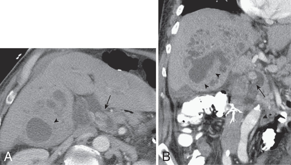
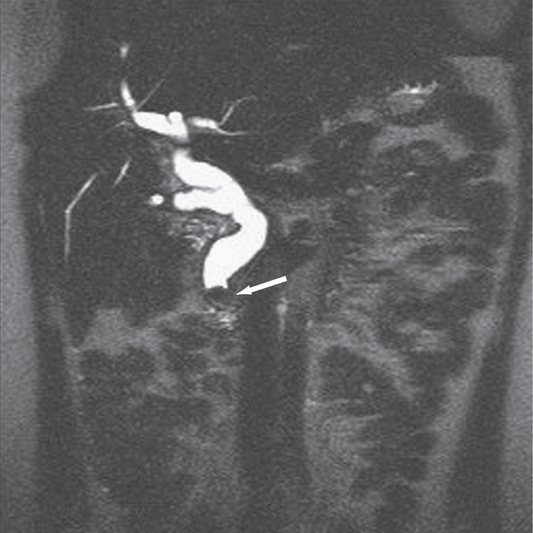
  • Other imaging modalities such as helical computed tomography (CT) (Fig. E2), magnetic resonance cholangiopancreatography (MRCP, Fig. E3), CT cholangiography, and endoscopic ultrasound (EUS) have improved performance characteristics for CBD stone detection; however, their use as first-line diagnostic tools is contingent on diagnostic uncertainty, patient factors, and availability.
  • Either MRCP or EUS should be used in intermediate-risk patients to confirm the diagnosis of choledocholithiasis and prompt further therapeutic management.
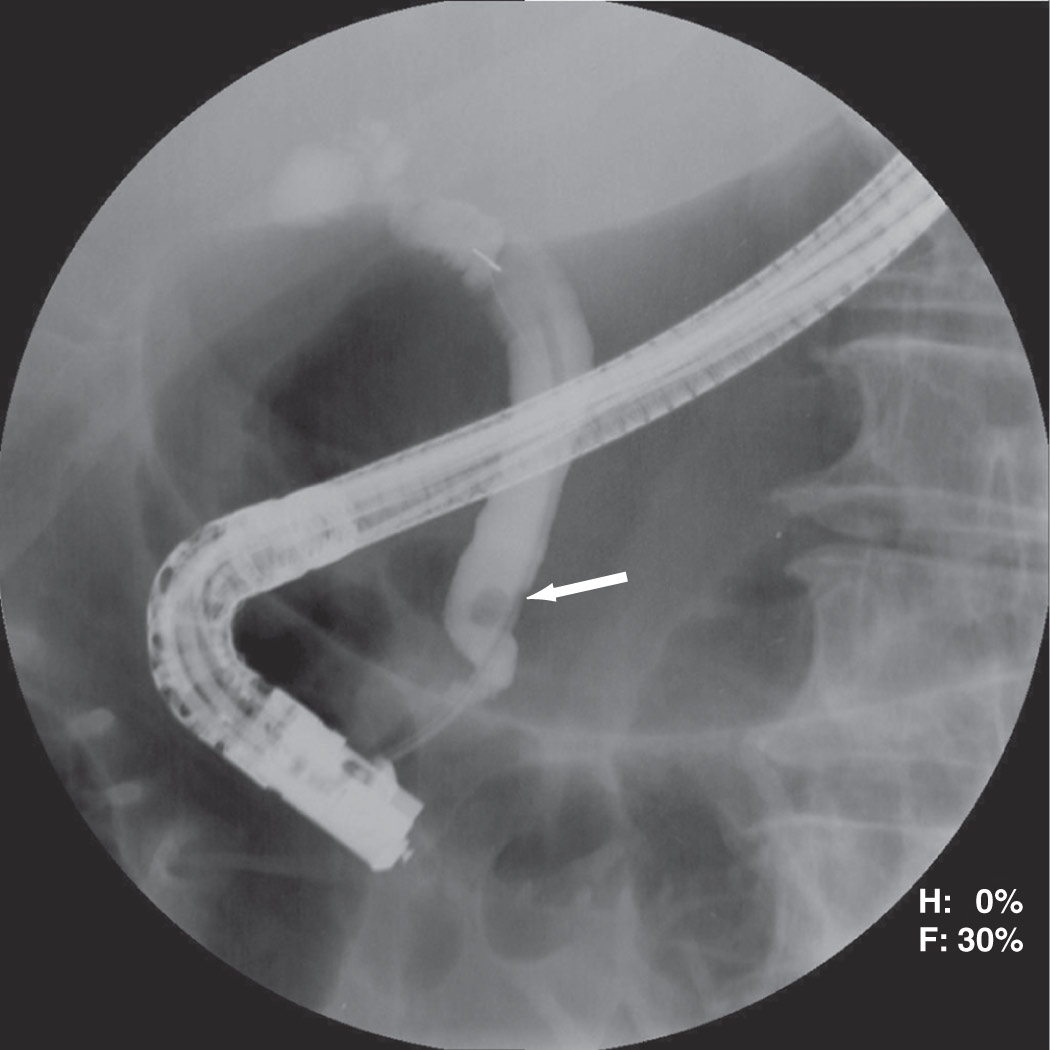
  • High-risk patients should proceed directly to endoscopic retrograde cholangiopancreatography (ERCP, Fig. E4) for diagnosis and treatment.

Figure E2 A 70-yr-old male with choledocholithiasis and a hepatic abscess.

Axial (A) and coronal (B) portal venous phase computed tomography images demonstrate large bile duct stones (arrows) with a focal intrahepatic fluid collection (arrowheads) consistent with the patient’s known pyogenic abscess.

(From Soto JA, Lucey BC: Emergency radiology: the requisites, ed 2, Philadelphia, 2017, Elsevier.)

Figure E3 Magnetic resonance cholangiopancreatography with choledocholithiasis.

The dilated common bile duct ends abruptly with a convex intraluminal filling defect (arrow) consistent with choledocholithiasis.

(From Townsend CM et al: Sabiston textbook of surgery, ed 21, St Louis, 2022, Elsevier.)

Figure E4 Endoscopic retrograde cholangiopancreatography (ERCP) with choledocholithiasis.

With retrograde injection of contrast material, a filling defect noted within the lumen of the common bile duct (arrow) identifies choledocholithiasis. ERCP can also be used to remove the stone through sphincterotomy and balloons or baskets.

(From Townsend CM et al: Sabiston textbook of surgery, ed 21, St Louis, 2022, Elsevier.)

Treatment

Acute General Rx

  • Presence of choledocholithiasis warrants treatment.
  • The mainstay is removal of CBD stones via ERCP and papillotomy either before or at the time of laparoscopic or open cholecystectomy.
  • Failure of ERCP to clear the biliary duct warrants biliary stenting for drainage as a temporary measure in the event of acute cholangitis.
  • Following biliary clearance with ERCP, it is generally recommended to proceed with subsequent cholecystectomy to prevent recurrence, which occurs in approximately 20% of patients.
  • The optimal timing of cholecystectomy after ERCP is still not clear, though performance of cholecystectomy within 24 h to 2 wk appears to be safe as earlier intervention is associated with a lower conversion of laparoscopic to open cholecystectomy.
  • ERCP without subsequent cholecystectomy can be considered definitive therapy in select high-risk patients with significant comorbidities as there is no statistical difference in mortality.
Chronic Rx

Patients with recurrent choledocholithiasis from cholesterol gallstones after cholecystectomy might be considered for chronic treatment with ursodeoxycholic acid to facilitate reduction of cholesterol saturation of bile.

Referral

  • Gastroenterology
  • General surgery
Related Content