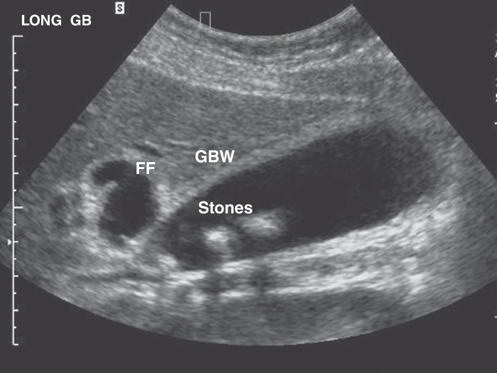
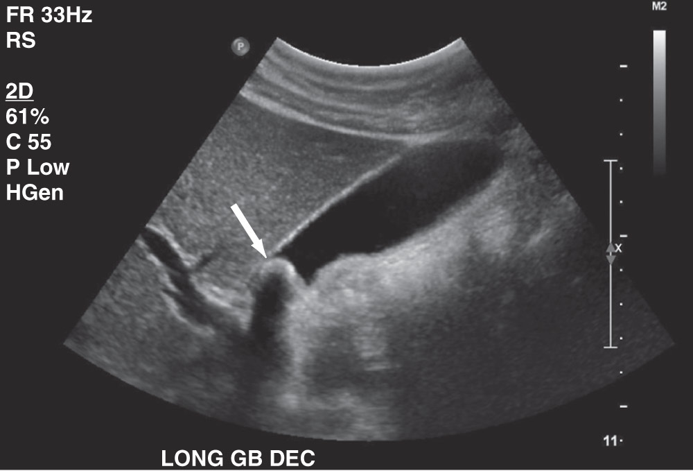
Author: Fred F. Ferri, MD


| ICD-10CM CODES | |||
| K80.80 | Other cholelithiasis without obstruction | ||
| K80.81 | Other cholelithiasis with obstruction | ||
| K91.86 | Retained cholelithiasis following cholecystectomy | ||
- •Gallstone disease can be found in 12% of the U.S. population. Of these, 2% to 3% (500,000 to 600,000) are treated with cholecystectomies each year.
- •Annual medical expenditures for gallbladder surgeries in the U.S. exceed $5 billion.
- •Incidence of gallbladder disease increases with age. Highest incidence is in the fifth and sixth decades. Predisposing factors for gallstones are female sex, pregnancy, age >40 yr, family history of gallstones, obesity, ileal disease, oral contraceptives, diabetes mellitus, rapid weight loss, estrogen replacement therapy.
- •Patients with gallstones have a 20% chance of developing biliary colic or its complications at the end of a 20-yr period. Significant predictors of gallstone-related events are large stone (>10 mm), presence of multiple stones, and female sex.
- •Physical examination is entirely normal unless patient is having biliary colic; 80% of gallstones are asymptomatic.
- •Typical symptoms of obstruction of the cystic duct include intermittent, severe, cramping pain affecting the right upper quadrant.
- •Pain occurs mostly at night and may radiate to the back or right shoulder. It can last from a few minutes to several hours.
- •Symptoms of gallstone disease and its complications are described in Table 2 and Fig. E1.
TABLE 2 Symptoms of Gallstone Disease and Its Complications
| Disease | Pathophysiology | Symptoms |
| Biliary colic | Transient gallstone impaction at the cystic duct or ampulla of Vater | Intermittent right upper quadrant pain associated with nausea or vomiting. Pain in the epigastrium or radiating to the right scapular tip. Episodes last 30 min to several hours with days or months between episodes. |
| Acute cholecystitis | Inflammation of the gallbladder caused by obstruction of the cystic duct. May occur in the presence or absence of bacterial superinfection | Patients appear ill and cannot take deep breaths. They have constant pain that lasts 30-60 min and worsens with movement. Persistent common bile duct impaction usually promotes vomiting. Physical examination demonstrates right upper quadrant tenderness with voluntary guarding and a positive Murphy sign (arrest of inspiration during deep palpation over the gallbladder). |
| Emphysematous cholecystitis | Infection with gas-producing bacteria such as Escherichia coli, Clostridium perfringens, and anaerobic streptococci | Symptoms are similar to those with acute cholecystitis. Gas may be seen on abdominal plain films or CT. Male diabetics are most commonly affected. |
| Chronic cholecystitis | Persistent inflammation and fibrosis of the gallbladder with poor motor and absorptive function | Patients are usually asymptomatic but may report multiple previous attacks of colic. Porcelain gallbladder develops from chronic inflammation and may progress to carcinoma. |
| Acalculous cholecystitis | Probably related to biliary stasis in the setting of critical illness and altered gastrointestinal motility | Seen in patients with traumatic injuries, burns, and critical illness, as well as in those receiving total parenteral nutrition. The mortality for this disorder is twice as high as that for acute calculous cholecystitis. |
| Gallbladder perforation | Stones erode through an inflamed and necrotic gallbladder wall. Stones may travel into the peritoneal cavity or cause adhesions between nearby structures. Bile peritonitis may develop | More than half of patients with gallbladder perforation have fever and a palpable right upper quadrant mass. Mortality in these patients is 30%. |
CT, Computed tomography.
From Adams JG et al: Emergency medicine, clinical essentials, ed 2, Philadelphia, 2013, Elsevier.
Figure E1 Schematic depiction of the natural history and complications of gallstones.
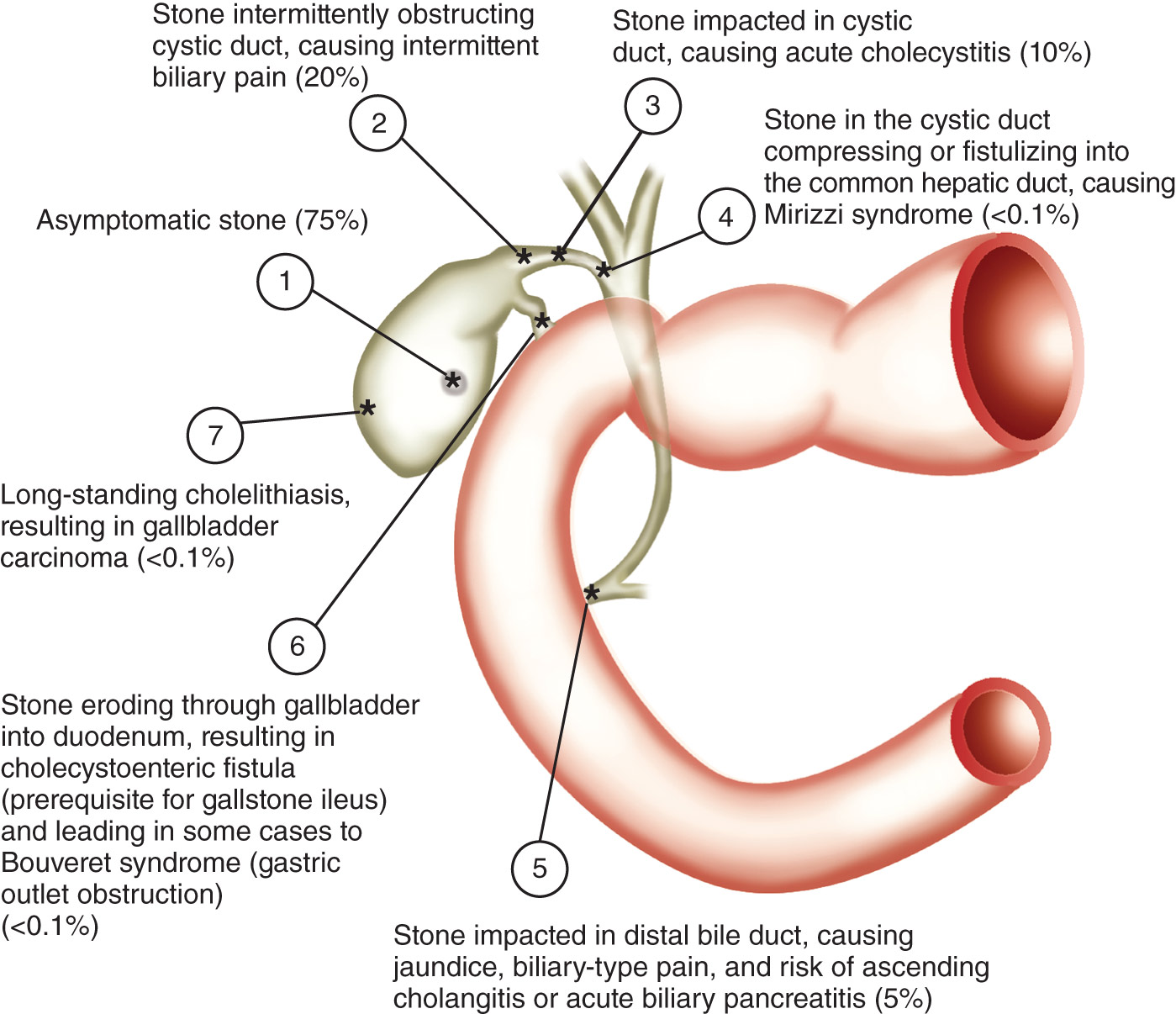
Percentages indicate approximate frequencies of complications that occur in persons with gallstones, based on natural history data. The most frequent outcome is for the patient with a stone to remain asymptomatic throughout life. (1) Asymptomatic or biliary pain, (2) acute cholecystitis, (3) cholangitis, and (5) pancreatitis are the most common complications. (4) Mirizzi syndrome, (6) cholecystoenteric fistula, (6) Bouveret syndrome, and (7) gallbladder cancer are uncommon. The sum of percentages is >100% because patients with acute cholecystitis generally have had prior episodes of biliary pain.
(From Feldman M et al: Sleisenger and Fordtran’s gastrointestinal and liver disease, ed 11, Philadelphia, 2021, Elsevier.)
TABLE 1 Pathophysiology, Clinical Manifestations, Diagnosis, and Treatment of Gallstone Disease
| Biliary pain | Acute Cholecystitis | Choledocholithiasis | Cholangitis | |
| Pathophysiology | ||||
| Symptoms | ||||
| Physical findings | ||||
| Laboratory findings |
|
|
| |
| Diagnostic studies | ||||
| Natural history | ||||
| Treatment | Elective laparoscopic cholecystectomy, possibly with IOC ERCP for stone removal or BD exploration if IOC shows stones | Laparoscopic cholecystectomy, possibly with IOC if feasible; otherwise open cholecystectomyBD exploration or ERCP for stone removal if IOC shows stones | Stone removal at the time of ERCP, followed in most cases by early laparoscopic cholecystectomy | Emergency ERCP with stone removal or at least biliary decompression Antibiotics to cover gram-negative and possibly anaerobic organisms and Enterococcus spp. Subsequent cholecystectomy |
BD, Bile duct; CT, computed tomography; ERCP, endoscopic retrograde cholangiopancreatography; EUS, endoscopic ultrasound; IOC, intraoperative cholangiography; MRC, magnetic resonance cholangiography; THC, transhepatic cholangiography; US, ultrasound; WBC, white blood cells.
From Feldman M et al: Sleisenger and Fordtran’s gastrointestinal and liver disease, ed 11, Philadelphia, 2021, Elsevier.
- •75% of gallstones contain cholesterol and are usually associated with obesity, female sex, and diabetes mellitus; mixed stones are most common (80%); pure cholesterol stones account for only 10% of stones and are the result of biliary supersaturation due to cholesterol secretion into the gallbladder and accelerated cholesterol nucleation and crystallization.
- •25% of gallstones are pigment stones (bilirubin, calcium, and variable organic material) associated with hemolysis and cirrhosis. These tend to be black-pigmented stones that are refractory to medical therapy.
- •50% of mixed-type stones are radiopaque.