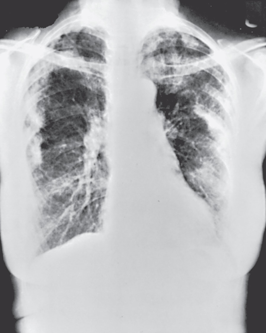
Author: Angela Suen, MD and Erica Farrand, MD

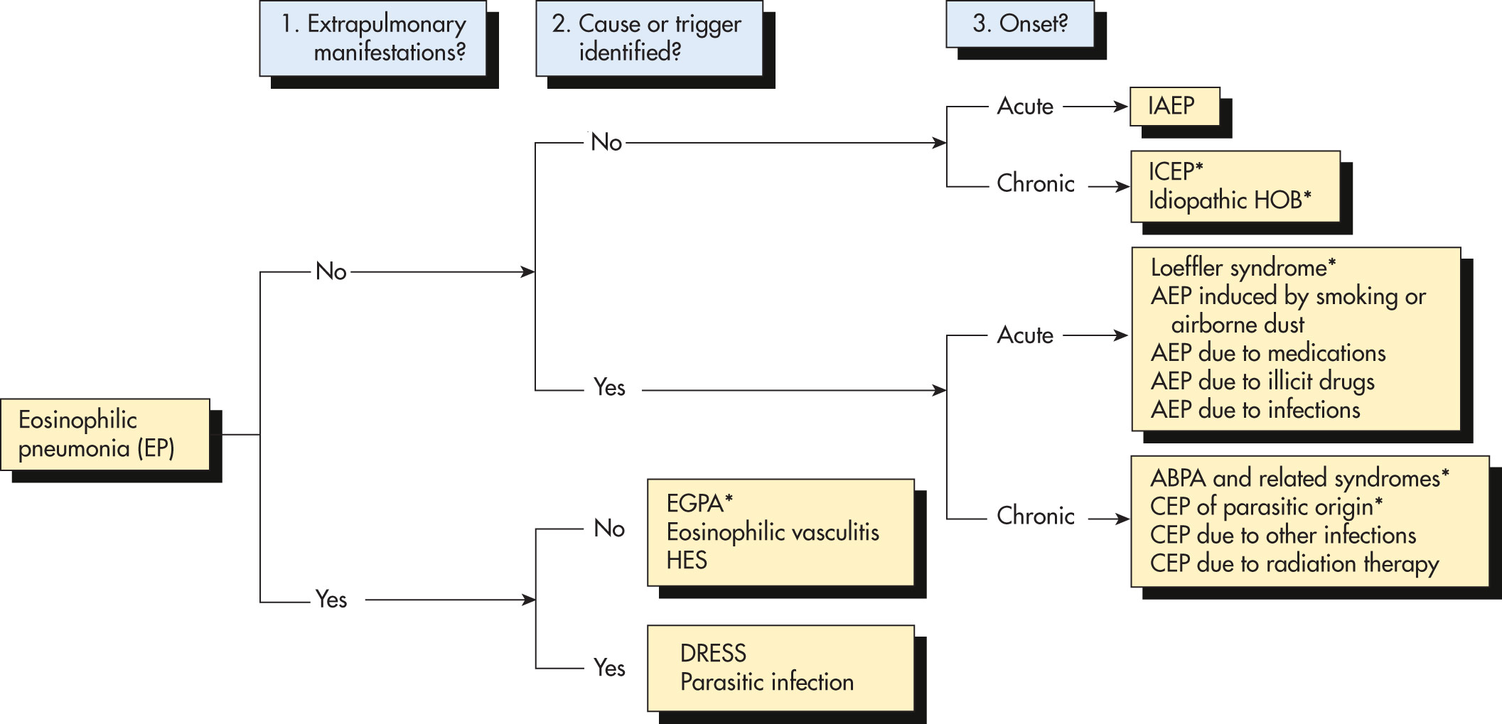
Eosinophilic pneumonias (EPs) are a group of heterogenous disorders characterized by eosinophilic infiltration of airways or lung parenchyma, and in some cases, peripheral blood eosinophilia. Although clinical and radiographic presentations are variable, EPs can generally be classified into lung-limited vs. systemic disorders.1
- EP
- Acute eosinophilic pneumonia
- Chronic eosinophilic pneumonia
- Loeffler syndrome
- Allergic bronchopulmonary aspergillosis
- Eosinophilic granulomatosis with polyangiitis (formerly Churg-Strauss syndrome)
- Idiopathic hypereosinophilic syndrome
- Parasite-induced (filariasis), fungal-induced, and drug-induced pulmonary eosinophilia
| ICD-10CM CODES | |||
| J82.81-J82.82, J82.89 | Pulmonary eosinophilia, not elsewhere classified | ||
| J70.2-J70.4 | Pulmonary eosinophilia due to drugs | ||
| B50-B83 | Pulmonary eosinophilia due to specified parasitic infection | ||
| M30-M36 | Pulmonary eosinophilia due to systemic connective tissue disorders | ||
| B44 | Pulmonary eosinophilia due to aspergillosis | ||
- •Fever, cough, and shortness of breath
- •Vary depending on the specific cause
Idiopathic acute eosinophilic pneumonia:
- •Associated with recent onset of tobacco smoking, inhalation of drugs, occupational exposures, exposure to World Trade Center dust, and military exposures in Middle East2
- •Acute-onset fever, cough, and dyspnea (<1 mo). Respiratory failure (PaO2<60 mm Hg) is common3,4
- •CT scan with patchy ground-glass and reticular opacities. Pleural effusions also common, which when sampled exhibit exudative characteristics with 10% to 50% eosinophilia
- •Diagnosis is made with bronchoalveolar lavage (BAL) >25% eosinophils and absence of infection or other causes of eosinophilic pneumonia
- •Often no peripheral blood eosinophilia
- •Steroids are the mainstay of therapy. Higher doses of intravenous methylprednisolone are used in patients with severe hypoxemic respiratory failure requiring mechanical ventilation, while oral prednisone 0.5 mg/kg/day tapered over 2 to 6 wk is used in less severe cases
- •Steroids lead to rapid improvement. Relapse is uncommon, and prognosis is excellent if promptly treated
Chronic eosinophilic pneumonia:
- •An idiopathic disorder that occurs in middle age.
- •More common in women and nonsmokers.
- •Associated with asthma in >50% of cases.
- •Gradual onset of productive cough, dyspnea, weight loss, night sweats, and fever over weeks to months.4
- •Chest x-rays (Fig. E1) typically show bilateral pulmonary opacities. "Photographic negative" pulmonary edema (peripheral or pleural-based opacities) is highly suggestive.
- •Diagnosis is made with BAL >25% eosinophils.5
- •Peripheral blood eosinophilia is almost always present. Immunoglobulin E (IgE) levels may also be elevated.
- •Pulmonary function tests are nonspecific and can show either restrictive or obstructive pattern. Thus they are more helpful in evaluating and monitoring degree of respiratory impairment rather than guiding diagnosis. Distinctive features of idiopathic chronic eosinophilic pneumonia and idiopathic acute eosinophilic pneumonia are summarized in Table E1.
- •Corticosteroids are the most effective first-line therapy at an initial dose of 0.5 mg/kg/day. Steroids are slowly tapered over the course of several months.
- •Case reports suggest inhaled steroids, omalizumab (monoclonal antibody to IgE), mepolizumab (monoclonal antibody to interleukin-5 [IL-5]), and benralizumab (monoclonal antibody to IL-5) may be helpful in patients with prolonged steroid dependence.6-10
- •Relapses are seen in over 50% of cases when corticosteroids are tapered or discontinued, and prolonged steroid courses are often required. Overall prognosis is typically excellent, and relapse does not portend a worse prognosis.
- •Due to transpulmonary passage of helminth larvae, most commonly Ascaris, hookworms, and Strongyloides, leading to transient pulmonary opacities and peripheral blood eosinophilia.
- •Symptoms are usually mild and self-limited. They include cough, dyspnea, wheezing, fever, and occasionally blood-tinged sputum that can contain Charcot-Leyden crystals.
- •When a parasitic infection is documented, appropriate anthelmintic drugs are indicated.
Figure E1 Idiopathic chronic eosinophilic pneumonia.
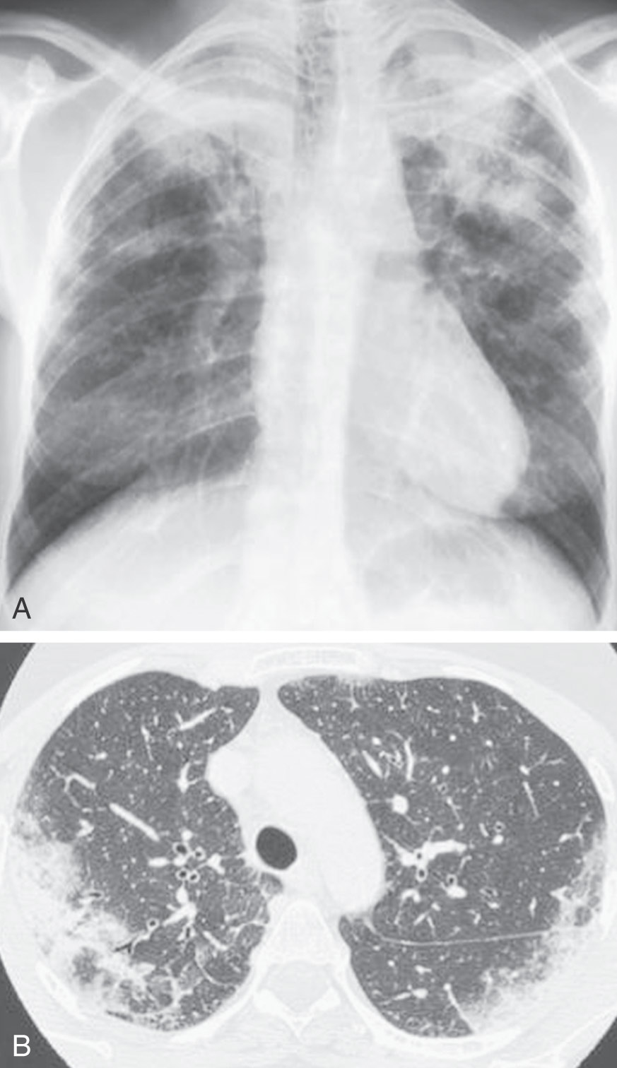
A, Chest radiograph of a patient with idiopathic chronic eosinophilic pneumonia shows bilateral alveolar opacities predominating in the upper lobes. B, Axial chest computed tomography scan of a patient with idiopathic chronic eosinophilic pneumonia shows bilateral symmetrical peripheral alveolar opacities.
(From Broaddus VC et al: Murray & Nadel’s textbook of respiratory medicine, ed 7, Philadelphia, 2022, Elsevier.)
TABLE E1 Distinctive Features of Idiopathic Chronic Eosinophilic Pneumonia and Idiopathic Acute Eosinophilic Pneumonia
BAL, Bronchoalveolar lavage; FiO2, fractional concentration of oxygen in inspired gas; IAEP, idiopathic acute eosinophilic pneumonia; ICEP, idiopathic chronic eosinophilic pneumonia; kPa, kilopascal; PaO2, arterial PO2.
From Broaddus VC et al: Murray & Nadel’s textbook of respiratory medicine, ed 7, Philadelphia, 2022, Elsevier.
Allergic bronchopulmonary aspergillosis (ABPA):
- •Occurs almost exclusively in patients with asthma or cystic fibrosis as a hypersensitivity reaction to Aspergillus fumigatus. Patients with chronic asthma, bronchiectasis, and pulmonary opacities should be evaluated for ABPA.11
- •Often manifests as severe, persistent asthma, resistant to treatment. Symptoms also include fever, myalgias, cough, wheezing, rhinosinusitis, expectoration of brown-black mucous plugs, and rarely hemoptysis.
- •Diagnostic criteria: Patients should meet the following criteria:12
- 1.History of asthma or cystic fibrosis
- 2.Obligatory criteria (both should be present):
- 3.At least two of the three minor criteria:
- a.Precipitating serum antibodies to Aspergillus
- b.Radiographic opacities consistent with ABPA (central bronchiectasis, endobronchial mucus impaction, recurrent upper/middle lobe predominant infiltrates)
- c.Peripheral blood eosinophilia >500 cells/microl in steroid naïve patients
- •Treatment: Systemic corticosteroids are the mainstay of treatment. Antifungals may provide additional benefit in modulating airway fungal burden and are recommended for use in patients with relapsed or glucocorticoid-dependent disease. If antifungal therapy is indicated, treat with oral itraconazole 200 mg twice daily, usually for at least 4 mo. Therapeutic drug monitoring is needed. Omalizumab (monoclonal antibody against IgE) or mepolizumab (monoclonal antibody against IL-5) may be beneficial in the treatment of ABPA in the setting of poorly controlled asthma.13
- •Improvement in symptoms and radiographic findings, and an IgE decrease by 25% indicates a response to therapy.
- •Development of bronchiectasis portends a worse prognosis. The disease has been characterized by a staging system. Patients do not necessarily proceed from one stage to another.
Stage I Acute phase Stage II Remission Stage III Exacerbation Stage IV Glucocorticoid-dependent ABPA Stage V End-stage (fibrotic) ABPA
Eosinophilic granulomatosis with polyangiitis (EGPA):
- •Formerly known as Churg-Strauss syndrome. Most commonly occurs in middle age.
- •Three phases of disease: Prodromal phase characterized by atopy, eosinophilic phase, and vasculitic phase.
- •During the vasculitic phase, eosinophilic granulomatous inflammation and necrotizing vasculitis of small to medium-size vessels lead to multiple organ involvement (lungs, heart, kidney, GI tract, skin, peripheral nervous system).
- •The American College of Rheumatology has established six classification criteria for EGPA: (1) Asthma, (2) peripheral blood eosinophilia, (3) mononeuropathy or polyneuropathy, (4) transient pulmonary opacities, (5) paranasal sinus abnormality, (6) biopsy showing extravascular accumulation of eosinophils. The presence of four or more has high sensitivity and specificity for the disease.14
- •35% to 40% of patients will have a positive antineutrophil cytoplasmic antibody (ANCA), and many will have an elevated IgE level.
- •Pulmonary function tests show variable airflow obstruction consistent with asthma.
- •Primary therapy is with systemic corticosteroids. Additional immunosuppression is used in patients with advanced/refractory disease or with flares when tapering steroids. Asthma should be treated concomitantly. Mepolizumab (monoclonal antibody against IL-5) is the only biologic approved for EGPA treatment.15
- •See chapter on Eosinophilic Granulomatosis With Polyangiitis.
- •A disease of persistently elevated peripheral eosinophils (>1500 cells/microl) with evidence of tissue deposition, resulting in end-organ damage.16
- •Can be due to myeloproliferative disorder, familial, or idiopathic.
- •Presentation depends on the organ systems involved, but can include fever, cough, weight loss, rash, wheezing, or central nervous system (CNS) abnormalities.
- •Lungs are affected in 25% of patients, and chest imaging may show opacities, consolidation, or lymphadenopathy.
- •Diagnosis is made by demonstrating persistently elevated peripheral eosinophilia (>1500 eosinophils/microl) with evidence of tissue deposition, resulting in end-organ damage. Cardiac disease in particular can be severe, and an echocardiogram should be done.
- •Asymptomatic patients may elect for close monitoring. Those with extremely high eosinophil counts, signs of leukocytosis, or end-organ damage should be treated urgently with corticosteroids. Risk assessment of Strongyloides infection before administration of steroids should be done.
- •Steroid-sparing agents, including hydroxyurea, methotrexate, and interferon alfa, can be considered in patients who relapse with steroid taper. Investigational studies for use of anti-IL5 antibodies (mepolizumab) are currently underway.
- •Patients with myeloproliferative disease associated hypereosinophilic syndrome with FIP1L1-PDGFRA fusion gene mutations typically have an aggressive course and are treated with imatinib.
Drug-induced eosinophilic pneumonia:
- •Most commonly associated with antibiotics, antiepileptics, and NSAIDs. Others include amiodarone, bleomycin, captopril, iodine, methotrexate, venlafaxine, and sulfa-containing drugs.
- •Can have several different clinical presentations, including simple pulmonary eosinophilia, chronic eosinophilic pneumonia, acute eosinophilic pneumonia, or drug reaction with eosinophilia and systemic symptoms (DRESS).17
- •Consider DRESS when pulmonary symptoms are also associated with skin eruption that involves more than 50% of the body surface area, fevers, liver function test abnormalities, and lymphadenopathy. Symptoms typically begin 2 to 6 wk after initiation of an offending agent.
- •Imaging findings are nonspecific and can include bilateral opacities.
- •Treatment includes removal of the offending agent. Severe cases sometimes require steroids.
Tropical pulmonary eosinophilia:
- •Clinical syndrome caused by immune hyper-responsiveness to filarial nematodes (roundworms). Majority of cases occur in endemic areas where filariasis occurs and most commonly in young adults.
- •Gradual onset of paroxysmal cough and bronchospasm associated with marked peripheral blood eosinophilia.
- •The diagnosis is confirmed with elevation in filarial antibody titers. High serum IgE levels are frequently observed.
- •Treatment is with diethylcarbamazine (DEC), and improvement is rapid in most cases. Relapse can occur in up to 20% of patients and is treated with a repeat course of DEC.