section name header

Basic Information ⬇

Author: Fred F. Ferri, MD

Figure E1 The metabolism and actions of vitamin D.
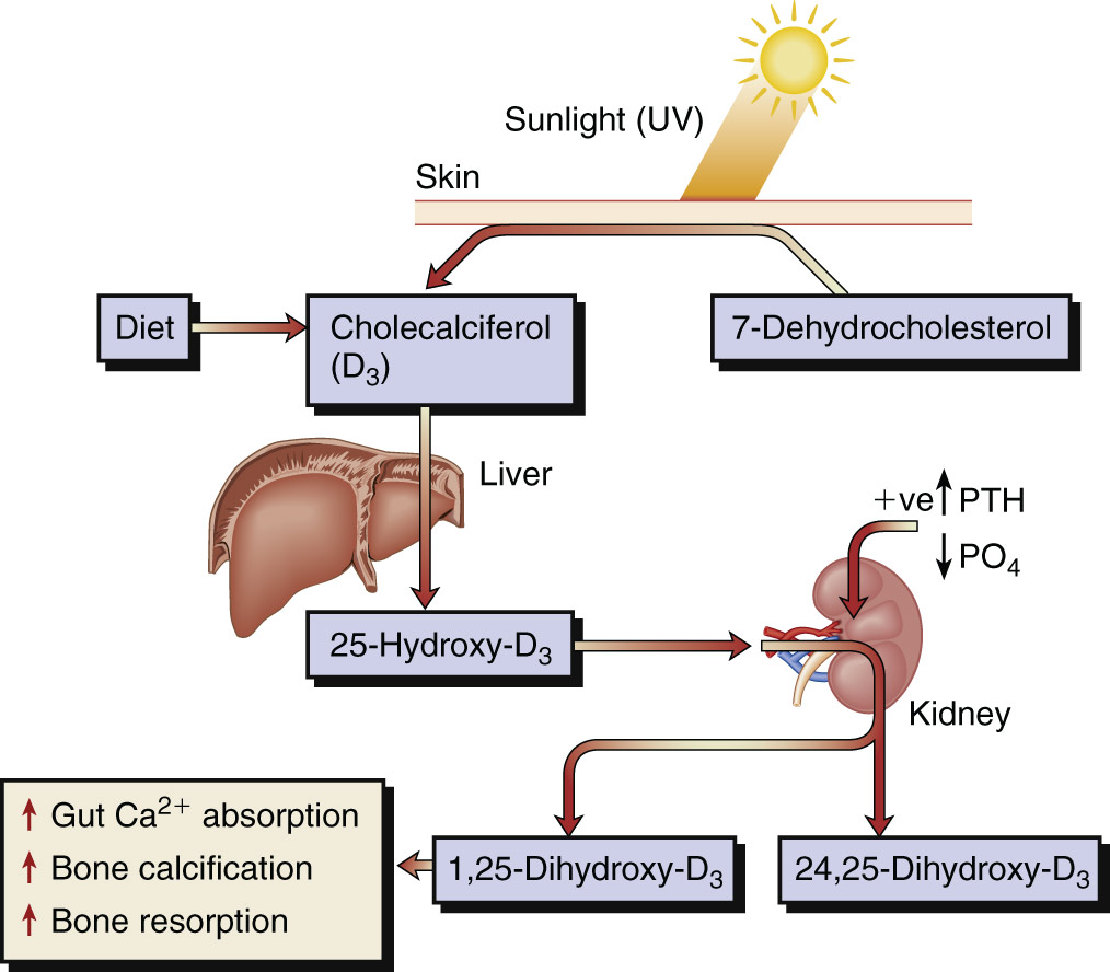
The primary source of vitamin D in humans is photoactivation in the skin of 7-dehydrocholesterol to cholecalciferol, which is then converted first in the liver to 25-hydroxyvitamin D and subsequently in the kidney to the much more active form, 1,25-dihydroxycholecalciferol (1,25[OH]2,D3). Regulation of the latter step is by parathyroid hormone (PTH), phosphate (PO4), and feedback inhibition by 1,25(OH)2,D3. This step can also occur in lymphomatous and sarcoid tissue, resulting in the hypercalcemia that may complicate these diseases. UV, Ultraviolet.

(From Ballinger A: Kumar & Clark’s essentials of clinical medicine, ed 6, Edinburgh, 2012, Saunders.)

Definition

Vitamin D deficiency is characterized by hypocalcemia and/or hypophosphatemia leading to impaired bone mineralization. It is classified as a serum 25-hydroxyvitamin D (25[OH]D) level of <20 ng/ml (50 nmol/L). This standard definition of vitamin D deficiency has been recently challenged, and some endocrinologists recommend a cutoff of 12 mg/ml for vitamin D deficiency. Vitamin D insufficiency is defined as a 25(OH)D between 12 and 20 ng/ml.

The consequences of vitamin D deficiency include:

  • •Bone disease (rickets, osteoporosis, low bone mass)
  • •May impair reproductive success
  • •Decrease the ability to combat infection (especially tuberculosis, influenza, viral infection)
  • •May induce or worsen autoimmune disorders
  • •May increase the incidence of death due to heart disease, inflammatory bowel disease, fracture, and cancer of the breast, colon, and prostate
  • •Subclinical vitamin D deficiency may occur in developed countries and be associated with increased fall risk and osteoporosis
Synonyms

  • The sunshine vitamin
  • The antirachitic factor
  • Cholecalciferol
ICD-10CM CODE
E55.9Vitamin D deficiency, unspecified
Epidemiology & Demographics
Incidence:

  • •Vitamin D insufficiency is very high among older adults and hospitalized and institutionalized people.
  • •Worldwide deficiency and insufficiency affect about 1 billion people.
  • •Children and young adults: 40% to 50% of preadolescent White girls, and Hispanic and Black adolescents, are vitamin D deficient.
Prevalence:

41.6% of adults (at least 20 yr old) have 25(OH)D levels <20 ng/dl.

Predominant Sex & Age:

  • •Decreased skin production of vitamin D with age
  • •Increased prevalence among darker-skinned individuals
Peak Incidence:

  • •In the U.S., 40% to 100% of the elderly are vitamin D deficient.
  • •Sixty percent of nursing home residents may be vitamin D deficient.
Risk Factors (Table 1):

  • •Age (due to decreased ability to produce D3)
  • •Sunshine-deficient areas (geographic location, living in higher latitudes)
  • •Dark-skinned individuals (melanin competes with vitamin D3 precursors for UV photons and thus decreases pre-D3 formation)
  • •Obese individuals
  • •Institutionalized individuals
  • •Pregnant and lactating women
  • •Use of sunscreen (sun radiation that causes skin cancer also produces pre-vitamin D3 in skin)
  • •Patients on certain medications that antagonize vitamin D action (phenobarbital, phenytoin)
  • •Intestinal resection
  • •Severe chronic liver diseases (such as cirrhosis)
  • •Kidney disease (e.g., nephritic syndrome)
  • •Sarcoidosis and lymphomas (increased catabolism of 25[OH]D to 1,25[OH]2D)
  • •Intestinal malabsorption disease (caused by celiac sprue, cystic fibrosis, Whipple disease)

TABLE 1 Classification of Rickets and Osteomalacia

  • Nutritional
    • Vitamin D deficiency
      • Dietary deficiency
      • Deficient endogenous synthesis
    • Calcium deficiency
      • Dietary deficiency
  • Gastrointestinal diseases
    • Intestinal
      • Small intestine diseases with malabsorption
      • Partial or total gastrectomy
      • Obesity and intestinal bypass
    • Hepatobiliary
      • Cirrhosis
      • Biliary fistula
      • Biliary atresia
    • Pancreatic
      • Chronic pancreatic insufficiency
  • Disorders of vitamin D metabolism
    • Hereditary
      • Isolated 25-hydroxyvitamin D deficiency
      • Pseudovitamin D-deficiency rickets
      • Hereditary vitamin D-resistant rickets
      • CYP3A4 gain-of-function mutations
      • FGF23 excess
    • Acquired
      • Hypoparathyroidism
      • Anticonvulsants
      • Renal insufficiency
      • FGF23 excess
  • Hypophosphatemia
    • FGF23-dependent
      • Genetic
        • XLH, ADHR, ARHR1, ARHR2
        • FAM20C mutation (Raine syndrome)
        • Fibrous dysplasia
      • Acquired
        • Tumor-induced osteomalacia
    • Non-FGF23-dependent
      • Genetic
        • Hereditary hypophosphatemic rickets with hypercalciuria
      • Dietary
        • Low phosphate intake with gastrointestinal disease
        • Alcohol use disorder
    • Hyperparathyroidism
  • Miscellaneous causes of impaired mineralization

  • Acidosis
    • Distal renal tubular acidosis
  • General renal tubular disorders (Fanconi syndrome)
    • Primary renal
    • Systemic disorder with associated tubular dysfunction
  • Chronic kidney disease
  • Aluminum, fluoride, and heavy metal intoxication
  • Hypophosphatasia
  • Osteopetrosis
  • Fibrogenesis imperfecta ossium

From Robertson RP et al: DeGroot’s endocrinology, basic science and clinical practice, ed 8, Philadelphia, 2023, Elsevier.

Physical Findings & Clinical Presentation

  • •Clinical presentation of vitamin D deficiency is dependent on the duration and severity of deficiency
  • •Most patients with mild to moderate vitamin D deficiency are asymptomatic
  • •Severe deficiency may lead to rickets (in children), osteomalacia (in adults), bone demineralization, hypokalemia, and phosphaturia
  • •Mild deficiency can lead to hypocalcemia and hyperparathyroidism
  • •Rickets (Tables 2 and 3): Seen in children; caused by defective mineralization in the skeleton (Figs. E2, E3)
    1. 1.Bowing of the legs
    2. 2.Leg bone pain
    3. 3.Delayed growth
    4. 4.Seizure due to hypocalcemia
  • •Osteomalacia: Seen in adults with severe and prolonged vitamin D deficiency
    1. 1.Periosteal bone pain (best detected by putting firm pressure on tibia or sternal bones)
    2. 2.Proximal muscle weakness
    3. 3.Chronic muscle aches/pain
  • •Fracture with very minimal trauma (brittle and easily broken bones)
  • •Severe hypocalcemia: Especially in late vitamin D deficiency leading to seizure tetany
  • •Hypophosphatemia
  • •Paresthesia
  • •Tetany
  • •Muscle cramps

TABLE 2 Causes of Rickets

Vitamin D Disorders
Nutritional vitamin D deficiency
Congenital vitamin D deficiency
Secondary vitamin D deficiency
Malabsorption
Increased degradation
Decreased liver 25-hydroxylase
Vitamin D-dependent rickets types 1A and 1B
Vitamin D-dependent rickets types 2A and 2B
Chronic kidney disease
Calcium Deficiency
Low intake
Diet
Premature infants (rickets of prematurity)
Malabsorption
Primary disease
Dietary inhibitors of calcium absorption
Phosphorus Deficiency
Inadequate intake
Premature infants (rickets of prematurity)
Aluminum-containing antacids
Renal Losses
X-linked hypophosphatemic rickets*
Autosomal dominant hypophosphatemic rickets*
Autosomal recessive hypophosphatemic rickets types 1, 2, and 3*
Hereditary hypophosphatemic rickets with hypercalciuria
Hypophosphatemic rickets with nephrolithiasis and osteoporosis types 1 and 2
Overproduction of fibroblast growth factor-23
Tumor-induced rickets*
McCune-Albright syndrome*
Epidermal nevus syndrome (cutaneous skeletal hypophosphatemia syndrome)*
Neurofibromatosis*
Fanconi syndrome
Dent disease
Distal renal tubular acidosis

* Disorders secondary to excess fibroblast growth factor-23.

From Kliegman RM et al: Nelson textbook of pediatrics, ed 22, Philadelphia, 2025, Elsevier.

TABLE 3 Clinical Features of Rickets

General
Failure to thrive (malnutrition)
Listlessness
Protruding abdomen
Muscle weakness (especially proximal)
Hypocalcemic dilated cardiomyopathy
Fractures (pathologic, minimal trauma)
Increased intracranial pressure
Head
Craniotabes
Frontal bossing
Delayed fontanel closure (usually closed by 2 yr)
Delayed dentition
No incisors by age 10 mo
No molars by age 18 mo
Caries
Craniosynostosis
Chest
Rachitic rosary
Harrison groove
Respiratory infections and atelectasis*
Back
Scoliosis
Kyphosis
Lordosis
Extremities
Enlargement of wrists and ankles
Valgus or varus deformities
Windswept deformity (valgus deformity of one leg with varus deformity of other leg)
Anterior bowing of tibia and femur
Coxa vara
Leg pain
Hypocalcemic Symptoms†
Tetany
Seizures
Stridor caused by laryngeal spasm

* These features are most frequently associated with vitamin D deficiency disorders.

† These symptoms develop only in children with disorders that produce hypocalcemia.

From Kliegman RM et al: Nelson textbook of pediatrics, ed 22, Philadelphia, 2025, Elsevier.

Figure E2 Radiographs of a Child with Vitamin D Deficiency Rickets, Demonstrating Bowing of the Femurs and Tibias (A) and Widened, Frayed, Demineralized Epiphyseal Plates (B and C)
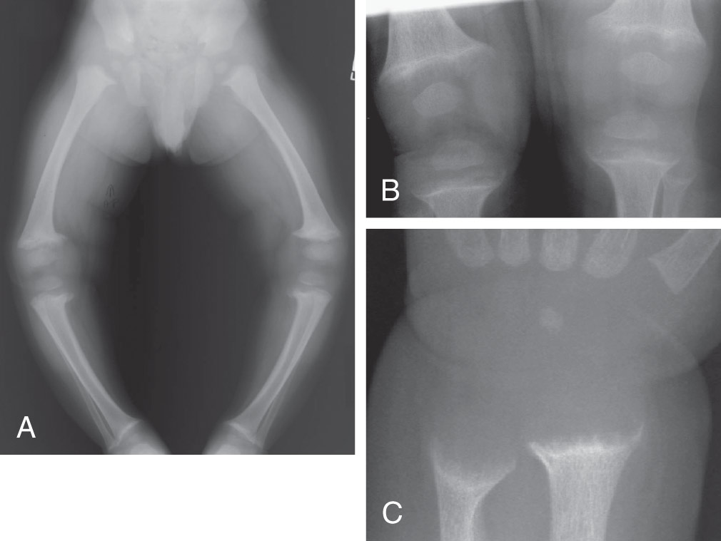
(Copyright of MF Holick, 2007. Reproduced with permission. In Hochberg MC et al: Rheumatology, ed 8, Philadelphia, 2023, Elsevier.)
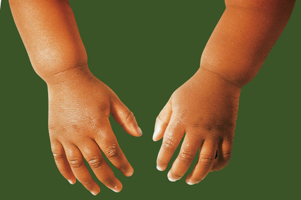
Figure E3 Hands and Forearms of a Young Child with Rickets Show Prominence above the Wrist, Resulting from Flaring and Poor Mineralization of Lower End of the Radius and Ulna

(From Bullough PG: Orthopaedic pathology, ed 5, St Louis, 2010, Mosby, Fig 8-31.)

Etiology

  • •Inadequate exposure to sunlight, such as:
    1. 1.During winter
    2. 2.In nursing home and health care institution residents
    3. 3.With excessive use of sunscreen
  • •Medications: Individuals on certain medications, such as phenobarbital, phenytoin, and rifampin (antagonize vitamin D action/increase vitamin D catabolism)
  • •Diseases and disease states:
    1. 1.Diseases causing vitamin D malabsorption:
      1. a.Cystic fibrosis
      2. b.Whipple disease
      3. c.Celiac sprue
    2. 2.Diseases increasing vitamin D catabolism:
      1. a.Lymphoma
      2. b.Sarcoidosis
    3. 3.Intestinal resection
    4. 4.Decreased 25(OH)D production:
      1. a.Kidney disease
      2. b.Liver cirrhosis

Diagnosis ⬆ ⬇

Differential Diagnosis

  • •Arthritis
  • •Fibromyalgia
Workup

  • •Population-wide screening for vitamin D deficiency is not recommended because evidence to support this practice is lacking. Appropriate to screen high-risk individuals.
  • •Screening is needed for individuals at risk (osteoporosis, history of falls, obese persons, pregnant and lactating women, diseases causing vitamin D malabsorption, Blacks). Workup involves blood and urine tests as well as radiography, as outlined in the next section.
Laboratory Tests (Table 4

  • •Serum 25(OH)D: This is the best test to determine vitamin D status.
  • •Parathyroid hormone (PTH): Increased levels in vitamin D insufficiency. It is a marker of vitamin D insufficiency.
  • •Increased (serum or bone) alkaline phosphatase.
  • •Decreased 24-h urine calcium (patient should not be on a thiazide).
  • •In patients at risk for osteomalacia [s-25(OH)D is less than 10 ng/ml], check calcium, Ph, alkaline phosphatase, PTH, basic metabolic panel, and tissue transglutaminase antibodies.

TABLE 4 Laboratory Tests

SERUMURINE
CalciumPhosphorusAlkaline PhosphataseCalcium
OsteoporosisNNNN
Hyperparathyroidism
Primary↑N or ↑N or ↑
SecondaryN or ↑↑↑↓
Tertiary↑N or ↓N or ↑N or ↑
Hypoparathyroidism↓↑N↓
Pseudohypoparathyroidism↓↑N↓
Rickets/osteomalacia
Vit D deficient↓↓↑
Vit D refractoryN↓↑↓
HypophosphatasiaN or ↑N↓N or ↑

Vit, Vitamin.

From Grant LA: Grainger & Allison’s diagnostic radiology essentials, ed 2, Philadelphia, 2019, Elsevier.

Imaging Studies

  • •Radiographs may show:
    1. 1.Pseudofractures of the pelvis, femur, metatarsals
    2. 2.Nontraumatic fractures
  • •Bone density:
    1. 1.Decreased bone mineral density (osteopenia or osteoporosis). Note that bone mineral density is not routinely performed in patients whose only risk factor is decreased Vitamin D levels.

Treatment ⬆ ⬇

Nonpharmacologic Therapy

  • •Natural sources of vitamin D. These include:
    1. 1.Exposure to sunlight. A mild sunburn is equivalent to consuming 10,000 to 25,000 IU of dietary vitamin D.
    2. 2.Dietary sources are not enough to meet daily requirements. Oily fish such as salmon, cod, and mackerel are rich sources of vitamin D3.
  • •Foods fortified with vitamin D
    1. 1.Mainly fortified dairy products
    2. 2.Fortified orange juice
Acute General Rx

  • •Treating deficiency (general population): Cholecalciferol (vitamin D3), when available, is preferred for vitamin D supplementation
    1. 1.50,000 IU (1250 mg) of vitamin D every wk for 8 wk, or
    2. 2.5000 IU (125 mg) daily to achieve a serum level of 25(OH)D of at least 30 ng/ml
  • •Maintenance measures after treatment (general population): 1500 to 2000 IU daily
  • •Treating deficiency (obese patients, patients with malabsorption syndromes, or those taking certain medications, as indicated earlier)
    1. 1.10,000 IU daily maintenance dose is recommended once s-25(OH)D level exceeds 30 ng/ml.
    2. 2.After treating deficiency, recheck 25(OH)D in 12 to 16 wk.
    3. 3.Maintenance measures after treatment (obese patients, patients with malabsorption syndromes, or those taking certain medications, as indicated earlier): 3000 to 6000 IU daily.
    4. 4.If deficiency persists after several attempts at treatment, try UV B light therapy.
Referral

Referral to an endocrinologist is recommended if there is no response to treatment.

Pearls & Considerations ⬆ ⬇

BOX E1 Symptoms of Vitamin D Toxicity

  • •Hypercalciuria and/or kidney stones/nephrocalcinosis
  • •Hypercalcemia and/or hyperphosphatemia
  • •Polyuria and polydipsia
  • •Decalcification of bone
  • •Ectopic calcification of soft tissues (kidney and lung)
  • •Nausea, vomiting, anorexia, or constipation
  • •Headache
  • •Hypertension

From Robertson RP et al: DeGroot’s endocrinology, basic science and clinical practice, ed 8, Philadelphia, 2023, Elsevier.

TABLE E5 Serum Concentrations of 25OHD

Normal Fluctuation According to
  • •Dietary intake (+)a
  • •Sun (ultraviolet [UV] light) exposure (+) influenced by seasonal lifestyle and cultural habits
  • •Age (–)
  • •Skin pigmentation (–)
  • •Latitude (–)
  • •Sunscreen use (–)
Increased 25OHD Concentration
  • •Exposure to pharmaceutical vitamin Db
  • •Excess exposure to nutritional vitamin D
  • •Excess exposure to UV light
  • •CYP24A1 deficiency
Decreased 25OHD Concentration
  • •Genetic or acquired deficiency of CYP2R1
  • •Genetic or acquired excess of CYP4A3
  • •Combined deficiency of access/exposure to nutritional vitamin D and UV light
    1. 1.Major risk groups include:
    2. 2.Infants, especially when born in late winter
    3. 3.Women and children of immigrants with pigmented skin living in temperate climates
    4. 4.Elderly individuals with limited mobility
    5. 5.Subset of the population with low exposure to sunlight because of socioeconomic, religious, or cultural reasons
  • •Decreased intestinal absorption of vitamin D associated with fat malabsorption (e.g., associated with biliary cirrhosis)
  • •Short bowel syndrome
  • •Exocrine pancreas insufficiency
  • •Gluten enteropathy
  • •Increased loss or catabolism of vitamin D
  • •Nephrotic syndrome
  • •Chronic liver P450 activation by drugs (e.g., barbiturates or antiepileptic drugs)
  • •Low calcium intake or absorption

a Positive or negative effects are indicated by + or –, respectively.

b Vitamin D toxicity with hypercalciuria, hypercalcemia, nephrocalcinosis, kidney stones, metastatic calcification, etc., is only observed if 25OHD concentrations exceed 100 ng/ml. Without access to pharmaceutical vitamin D, it is therefore uncommon to develop clinical vitamin D toxicity.

From Robertson RP et al: DeGroot’s endocrinology, basic science and clinical practice, ed 8, Philadelphia, 2023, Elsevier.

TABLE E6 Serum Concentration of 1,25(OH)2 D

Decreased ConcentrationsIncreased Concentrations
Substrate DeficiencyaSubstrate Excessb
(e.g., nutritional rickets, intestinal malabsorption)
25OHD-1α-Hydroxylase25OHD-1α-Hydroxylase
Enzyme DeficiencyEnzyme Excess
Inborn: Vitamin D-dependent ricketsPrimary or tertiary hyperparathyroidism
Organic: Renal insufficiency or anephric patientsHypothyroidism
Functional:Glucocorticoid excess
HypoparathyroidismAcromegaly
PseudohypoparathyroidismGranulomatous diseases
HypomagnesemiaIdiopathic hypercalciuria
Tumoral osteomalaciaHypophosphatemic rickets type 2 (+hypercalciuria)
Hypercalcemia of malignancyPregnancy
HyperthyroidismNutritional calcium deficiency
Morbus Addison (acute)Williams syndrome
Severe insulin deficiency
X-linked hypophosphatemia
Rhabdomyolysis
Tumoral calcinosis
DBP DeficiencyDBP Excess
FetusPregnancy
Nephrotic syndromeOral estrogen use
Liver cirrhosis
Genetic mutation
End-Organ Resistance
True vitamin D resistance (so-called vitamin D-dependent rickets type 2)

DBP, Vitamin D-binding protein.

a In many cases of rickets or osteomalacia, 1,25(OH)2D concentrations are still measurable or even nearly normal. This may be as a result of recent (and insufficient) access to vitamin D after long-term vitamin D deficiency. Nevertheless, such concentrations are too low in comparison with the degree of secondary hyperparathyroidism. Regardless, 25(OH)D is a better marker for vitamin D deficiency than 1,25(OH)2D. A similar situation is observed in hypothyroidism, as the precursor hormone T4 is a better marker for clinical hypothyroidism than the real hormone, T3 .

b Vitamin D excess only increases serum l,25(OH)2D when renal function remains normal and/or parathyroid hormone secretion is elevated. Frequently, l,25(OH)2D levels are low or normal in vitamin D toxicity.

From Robertson RP et al: DeGroot’s endocrinology, basic science and clinical practice, ed 8, Philadelphia, 2023, Elsevier.

Prevention

  • •Food fortification with vitamin D2 or vitamin D3
  • •Adequate sun exposure, for example, exposure in the middle of the day (between 10:00 A.M. and 3:00 P.M.)
  • •Use vitamin D3 for supplementation when available
  • •Vitamin D supplementation (per the Endocrine Society):
    1. 1.Infants (age range 1 to 12 mo) require at least 400 IU/day of vitamin D
    2. 2.Children (age range 1 to 18 yr) require 600 IU/day of vitamin D
    3. 3.Adult supplementation (adults 19 to 70 yr): 600 IU of vitamin D daily
    4. 4.Adult supplementation (persons ≥70 yr): 800 IU of vitamin D daily
    5. 5.Exceptions: Pregnant or lactating women, obese persons, and patients on antiseizure medications, steroids, antifungals, and AIDS medications should be given 2× to 3× more vitamin D
    6. 6.To reduce the risk of fracture and falls, the American Geriatric Society recommends a daily intake of at least 1000 IU and the National Osteoporosis Foundation 800 to 1000 IU in adults 65 yr or older
Screening

Routine screening for low-risk adults is not recommended. Screening is recommended only for individuals at high risk for vitamin D deficiency such as Blacks and Hispanics, obese individuals (body mass index >30 kg/m2), patients with osteoporosis, the elderly, and patients with certain chronic diseases (see "Risk Factors"). According to the U.S. Preventive Services Task Force, current evidence is insufficient to assess the balance of benefits and harms of screening for vitamin D deficiency in asymptomatic adults.

Related Content

Reference(s) ⬆

  1. Appel LJ : The effects of four doses of vitamin D supplements on falls in older adults: a responsive-adaptive, randomized clinical trialAnn Intern Med. 174(2):145-156, 2021.
  2. Waterhouse M : The effect of monthly vitamin D supplementation on fractures: a tertiary outcome from the population-based double-blind randomised, placebo-controlled D-Health trialLancet Diabetes Endocrinol. 11(5):324-332, 2023.
  3. Neale RE : The D-health trial: a randomized controlled trial of the effect of vitamin D on mortalityLancet Diabetes Endocrinol. 10(2):120-128, 2022.
  4. Hahn J : Vitamin D and marine omega 3 fatty acid supplementation and incident autoimmune disease: VITAL randomized controlled trialBMJ. 376:e066452, 2022.