section name header

Basic Information ⬇

Author: Fred F. Ferri, MD

Definition

Vitamins are organic compounds that cannot be synthesized by humans but are required as nutrients in minute amounts for normal metabolism. Vitamins have several different functions: They may regulate cell growth and differentiation, as catalysts, as antioxidants, and as coenzymes. Vitamins are classified as either fat soluble (vitamins A, D, E, K) or water soluble (B group of vitamins and C). Deficiency of most vitamins is rare in Western countries. Certain groups may be prone to vitamin deficiency, and these are discussed here. Vitamin D deficiency is discussed in a separate topic.

Synonyms

  • Hypovitaminosis
  • Vitamin A: Retinol
  • Vitamin E: Alpha tocopherol
  • Vitamin K: Phytonadione or menadiol
  • Vitamin B1: Thiamine
  • Vitamin B2: Riboflavin
  • Niacin: Vitamin B3; nicotinic acid
  • Vitamin B5: Pantothenic acid
  • Vitamin B6: Pyridoxine; pyridoxal phosphate
  • Folic acid: Vitamin B9; folate
  • Vitamin B12: Cyanocobalamin
  • Vitamin C: Ascorbic acid
ICD-10CM CODES
E50Vitamin A deficiency
E51Thiamine deficiency
E53Deficiency of other B group vitamins
E55Vitamin D deficiency
E56Other vitamin deficiencies
E56.0Deficiency of vitamin E
E56.1Deficiency of vitamin K
E53.0Riboflavin deficiency
E52Niacin deficiency [pellagra]
E53.1Pyridoxine deficiency
E53.8Deficiency of other specified B group vitamins
E54Ascorbic acid deficiency
Epidemiology & Demographics

Deficiency can occur in all age groups but is most common in the elderly.

  • •Vitamin A deficiency: Affects 250 million preschool children worldwide.
  • •Vitamin E deficiency: Deficiency is rare in humans. Usually occurs in individuals with severe protein-energy malnutrition.
  • •Vitamin K deficiency: Varies by geographic regions; no race predilection; affects both sexes equally. Encountered often in infants. In normal healthy adults, 8% to 31% have vitamin K deficiency, but it rarely leads to significant bleeding.
  • •Vitamin B1 (thiamine) deficiency: Incidence is unknown; no sex, race, or age predilection. Deficiency is usually due to inadequate intake, especially if consuming diet made up of polished rice and grains.
  • •Vitamin B2 (riboflavin): More common than previously appreciated. Deficiency is referred to as ariboflavinosis.
  • •Vitamin B5 (pantothenic acid) deficiency: Rare, as it is present in all foods.
  • •Vitamin B12 (cobalamin) deficiency: Relatively common. Of patients with anemia, about 1% to 2% is due to B12 deficiency. Among patients with macrocytosis (mean corpuscular volume [MCV] >100) 18% to 20% is due to B12 deficiency. Occurs in all age groups but more common in the elderly. B12 deficiency due to pernicious anemia is common in Northern Europe.
  • •Vitamin B9 (folic acid) deficiency: Mandatory fortification started in 1998. Prevalence before fortification 16% and after 0.5%. Neural tube defect associated with low maternal folate status during pregnancy. Pregnant women and the elderly are at greatest risk of folic acid deficiency.
  • •Vitamin C (ascorbic acid) deficiency: Smokers and low-income persons are at increased risk. Vitamin C deficiency is associated with access to food and/or socioeconomic status. Prevalence varies worldwide, but the rate is about 7.1% in the U.S.
  • •Vitamin E: Vitamin E depends on the presence of pancreatic esterases and bile salts for its solubilization and absorption in the intestinal lumen. Neurologic symptoms of deficiency occur most commonly in patients with fat malabsorption. A reduced bile salt pool may be caused either by reduced hepatic excretion, as in congenital cholestasis, or by interruption of the enterohepatic reabsorption of bile, as in patients with extensive small-bowel resection. Pancreatic insufficiency contributes to malabsorption. Another setting is cystic fibrosis.

Fig. 1 shows environmental and nutritional factors in disease.
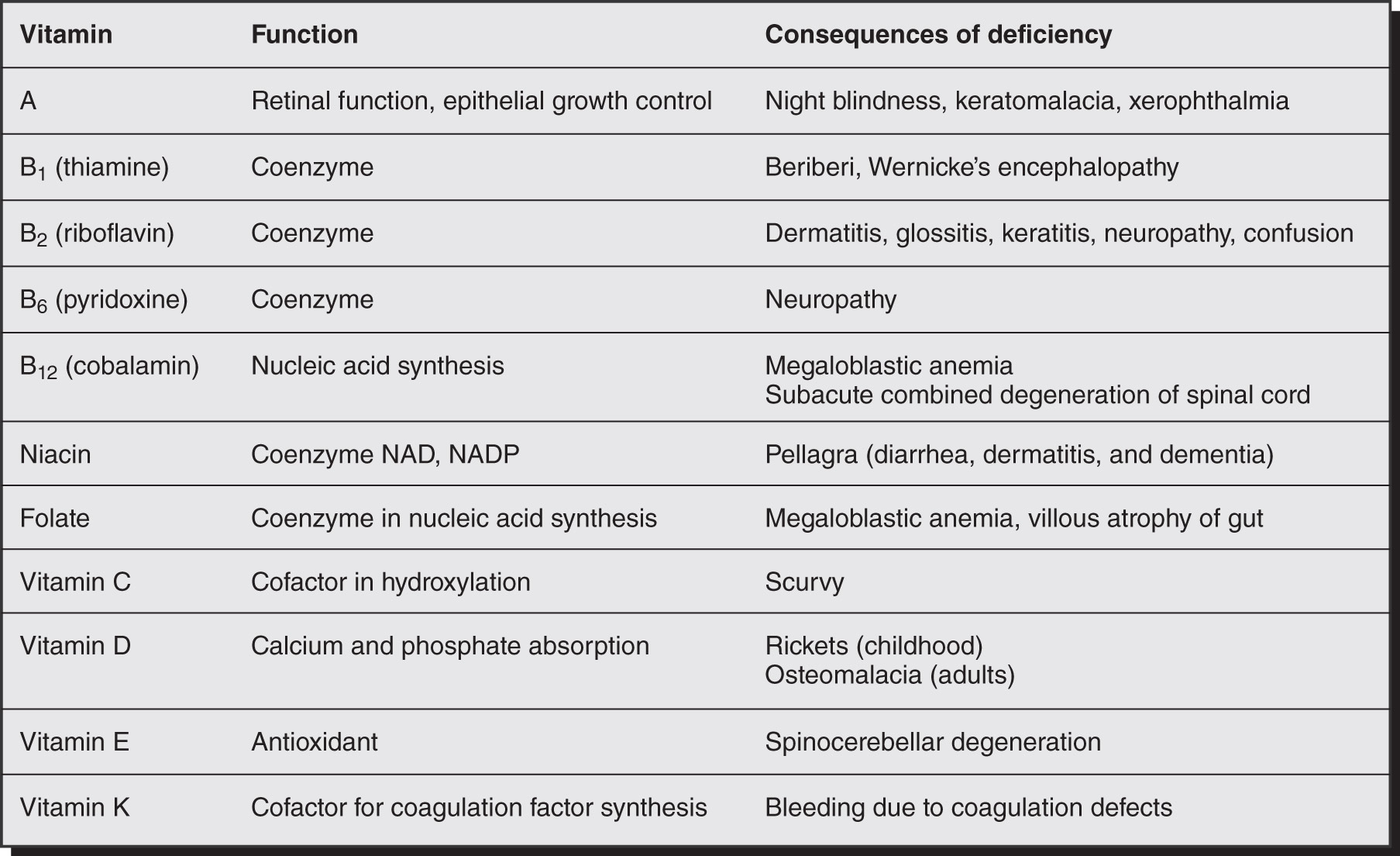
Figure 1 Environmental and nutritional factors in disease.

(From Stevens A: Core pathology, St Louis, 2009, Elsevier.)

Physical Findings & Clinical Presentation

  • •Vitamin A: Xerophthalmia, xerosis of the cornea, keratomalacia, Bitot spots (abnormal squamous cell proliferation and keratinization of the conjunctiva), nyctalopia (poor adaptation to darkness)/night blindness, poor bone growth, dry skin and hair, follicular hyperkeratosis (caused by blockage of hair follicles by keratin), pruritus, broken fingernails
  • •Vitamin K: Clinical manifestation usually occurs if hypoprothrombinemia is present. Major symptom is bleeding to minor trauma. Also can show easy bruisability, epistaxis, hematoma, gum bleeding, melena, hematuria, or splinter hemorrhage
  • •Vitamin E: Neuromuscular disorders (ataxia; hyporeflexia, peripheral neuropathy); bone weakness, hemolysis
  • •Vitamin B1 (thiamine): Beriberi, which has two subtypes (infantile and adult). Adult type is described below:
    1. 1.Dry beriberi (affecting the nervous system): Symmetric peripheral neuropathy (with sensory and motor impairments), Wernicke encephalopathy (nystagmus, ataxia, ophthalmoplegia, and confusion), Korsakoff syndrome (impaired short-term memory loss and confabulation but normal cognition)
    2. 2.Wet beriberi (affecting the cardiovascular system): Cardiomegaly, cardiomyopathy, heart failure, tachycardia, hypotension, chest pain, peripheral edema
    3. 3.Gastrointestinal (GI): Anorexia; constipation
  • •Vitamin B2 (riboflavin):
    1. 1.Cheilosis (chapping and fissure of the lip)
    2. 2.Glossitis (sore red tongue)
    3. 3.Oily, scaly rashes on nasolabial folds, eyelids, scrotum, labia majora
    4. 4.Red itchy eyes
    5. 5.Normocytic or normochromic anemia
    6. 6.Peripheral neuropathy
  • •Vitamin B3 (niacin):
    1. 1.Pellagra (4 Ds-diarrhea, dermatitis, dementia, and ultimately death)
    2. 2.Hyperpigmentation of sun-exposed skin
    3. 3."Raw beef" swollen and painful tongue
    4. 4.Deficiency can be seen in prolonged use of isoniazid, in carcinoid syndrome, and in Hartnup syndrome
  • •Vitamin B5 (pantothenic acid):
    1. 1.Deficiency is rare
    2. 2.Deficiency leads to "burning feet syndrome" (distal paresthesia and dysesthesia)
    3. 3.Anemia
    4. 4.GI symptoms
  • •Vitamin B6 (pyridoxine): Rare to see overt deficiency
    1. 1.Mild deficiency: Glossitis, cheilosis, impaired proprioception; sensory ataxia, confusion, depression
    2. 2.Severe deficiency: Seborrheic dermatitis, seizure, microcytic
  • •Vitamin B12 (cyanocobalamin):
    1. 1.Megaloblastic anemia (pernicious anemia)
    2. 2.Neurologic symptoms including peripheral neuropathy, ataxia (shuffling gait), paresthesia; subacute degeneration of the spinal cord (demyelination of the dorsal column, [Fig. E2]), visual disturbances due to optic atrophy
    3. 3.Glossitis and GI symptoms such as nausea, vomiting, and anorexia are also common
    4. 4.Patients may also have dementia/mental sluggishness, depression, and weakness
  • •Vitamin B9 (folic acid):
    1. 1.Patchy hyperpigmentation of skin (especially between fingers and toes) and mucous membranes
    2. 2.Moderate fever (temp <102° F; 38.9° C) despite the absence of infection
    3. 3.Neural tube defect
    4. 4.Angular stomatitis
    5. 5.Red, beefy, smooth, and shiny tongue
    6. 6.Megaloblastic anemia
  • •Vitamin C: Scurvy (bruising, petechiae, follicular hyperkeratosis, perifollicular hemorrhage [Fig. E3], corkscrew hairs), poor wound healing, fatigue, gingivitis/bleeding gums, weight loss, bone abnormalities (Fig. E4). Also, loss of teeth, abnormal nail (koilonychia and splint hemorrhages). Vitamin C deficiency may be associated with nonalcoholic fatty liver

Figure E2 Spinal cord in cobalamin deficiency.
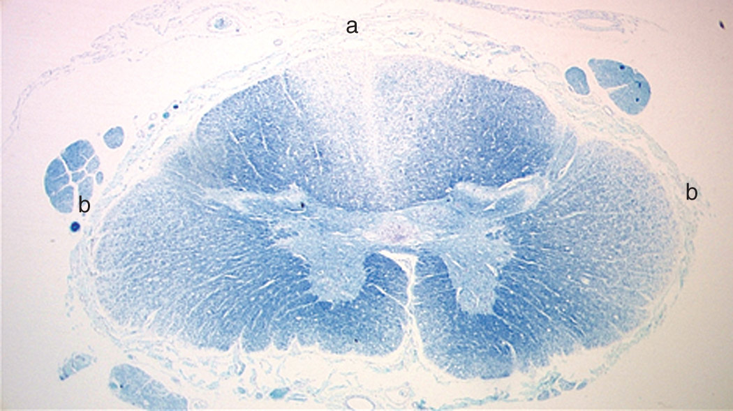
The cross-section of the spinal cord stained with Luxol blue shows demyelination of the dorsal columns (a) and early demyelination of the lateral columns (b).

(From Hoffman R et al: Hematology, basic principles and practice, ed 8, Philadelphia, 2023, Elsevier.)
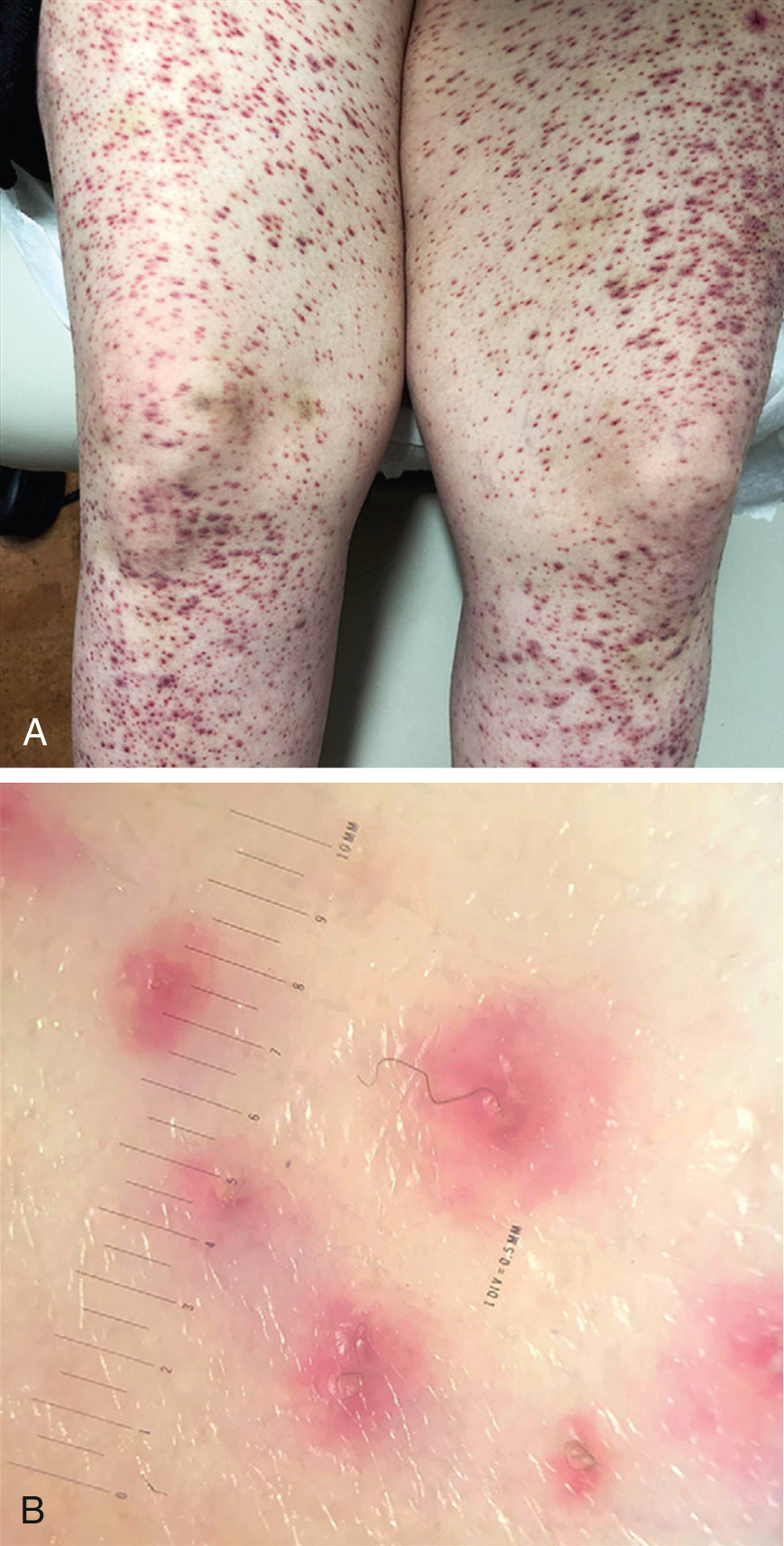
Figure E3 Scurvy.

A, Perifollicular hemorrhages. B, Corkscrew hairs with perifollicular hyperkeratotic papules.

(Modified from Lipner S: A classic case of scurvy, Lancet 392[10145]:431, 2018.)
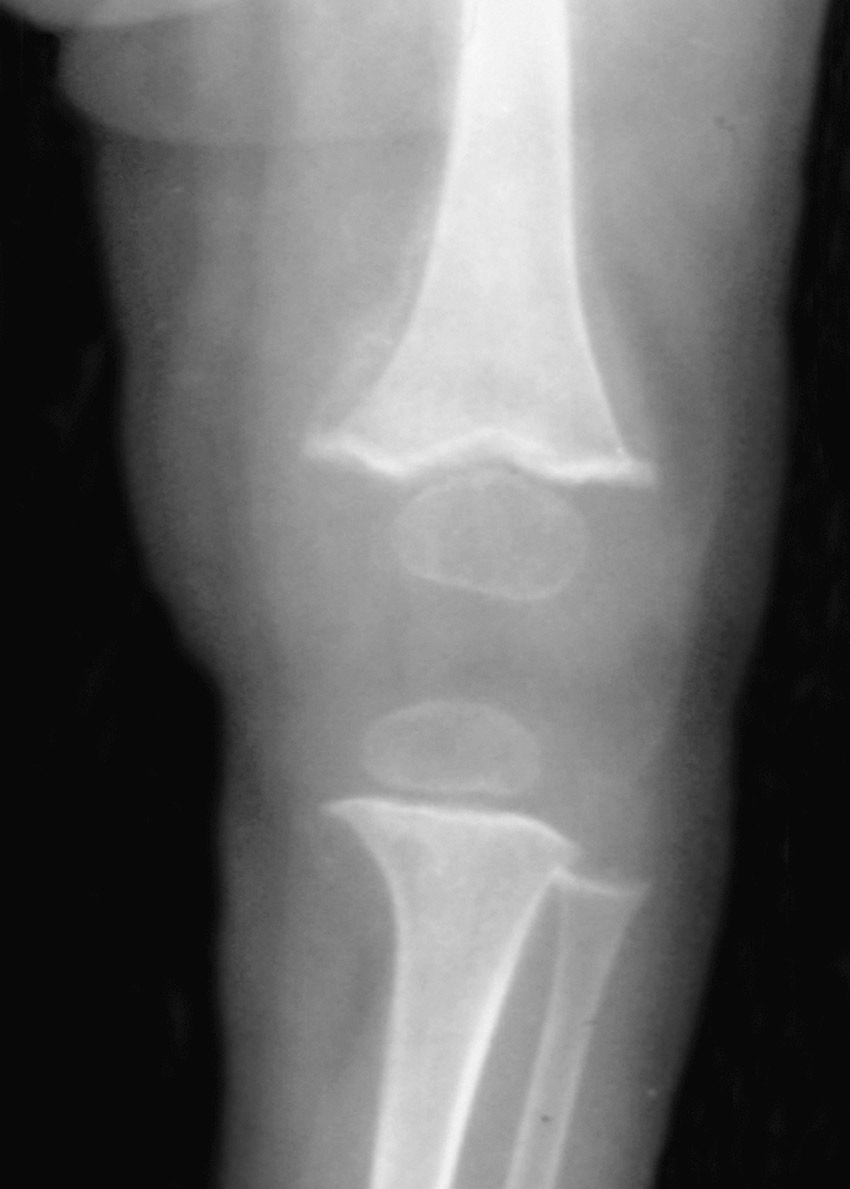
Figure E4 Scurvy.

The knee shows widened metaphyses with spurs and reduced bone density. The ossific centers have a typical "white pencil" outline. Faint periosteal reaction is visible in the distal femur secondary to periosteal hemorrhage. The end plate is still well defined, and the physis is not widened (compared with rickets). Fractures are rare.

(From Pope TL et al: Musculoskeletal imaging, ed 2, Philadelphia, 2014, Saunders.)

Etiology

  • •Fat-soluble vitamins (vitamins A, D, E, K):
    1. 1.Decreased ingestion, malnutrition, eating disorders
    2. 2.Diseases that affect fat absorption decrease the absorption of fat-soluble vitamins-for example, cystic fibrosis, celiac sprue, inflammatory bowel disease, cholestasis, hepatobiliary disease, small bowel surgery
    3. 3.Change in vitamin metabolism:
      1. a.Alcoholism
      2. b.Drugs such as cholestyramine, warfarin, anticonvulsants, antibiotics (e.g., cephalosporins)
      3. c.Chronic kidney disease
  • •Increased risk in:
    1. 1.Vegans
    2. 2.Recent immigrants
    3. 3.Refugees
    4. 4.Toddlers/preschoolers living below the poverty line
  • •Water-soluble vitamins (the B group of vitamins and vitamin C)-there are several etiologic factors, including:
    1. 1.Inadequate intake
    2. 2.Decreased absorption
    3. 3.Alcoholism
    4. 4.Pregnancy/lactation
    5. 5.Peritoneal dialysis
    6. 6.Medications (e.g., isoniazid, phenothiazines, tricyclic antidepressants, metformin [vitamin B12])
    7. 7.Malabsorption
    8. 8.Low income
    9. 9.Advanced age
  • •Vitamin B12 deficiency-caused by:
    1. 1.Insufficient dietary intake, as in strict vegans
    2. 2.Decreased absorption secondary to intrinsic factor deficiency, decreased intrinsic factor secretion, gastric atrophy, gastrectomy/gastric bypass
    3. 3.Terminal ileum disease such as celiac disease, enteritis, tropical sprue
  • •Folic acid deficiency:
    1. 1.Increased needs can lead to deficiency (e.g., pregnancy, lactation, malignancy)
    2. 2.Derangement of folate metabolism by:
      1. a.Medication (e.g., methotrexate)
      2. b.Disease (e.g., hypothyroidism)
      3. c.Increased excretion: As seen in alcoholics

Diagnosis ⬆ ⬇

Workup

Table 1 summarizes clinical clues in identifying vitamin deficiency. A stepwise approach to the diagnosis of cobalamin and folate deficiency is summarized in Table 2. False positive and false negative test results are explained in Table 3.

TABLE 1 Clinical Clues in Identifying Vitamin Deficiency

Clinical FeaturesCauses and DiagnosisTreatment and Notes
Vitamin A deficiency
  • Can take years to cause symptoms
  • Xerophthalmia causing night blindness and Bitot spots (conjunctival squamous cell proliferation and keratinization) is the earliest sign
  • Poor bone growth
  • Follicular hyperkeratosis
  • Impaired immune system
  • Conjunctival xerosis
  • Keratomalacia
  • Low dietary intake (preformed vitamin A is from animals; provitamin A is found in plants)
  • Diagnosis is made by measuring serum retinol levels
  • Vitamin A supplementation
  • Daily requirement (RDA) for adult males is 3000 IU and for females is 2300 IU
  • Vitamin A toxicity is related to chronic ingestion (≥25,000 IU/day); serum retinol levels are not helpful as vitamin A is stored in the liver
Vitamin B12 deficiency
  • Can take several years to show symptoms
  • Macrocytic anemia
  • Smooth tongue
  • In severe deficiency-subacute combined degeneration of the spinal cord
  • Peripheral sensory neuropathy affecting large and small fibers
  • Dementia
  • Low dietary intake
  • Pernicious anemia
  • Terminal ileum disease
  • Vitamin B12 supplementation
  • If both folate and vitamin B12 deficiency are present, you must replace vitamin B12 first to avoid subacute combined degeneration of the spinal cord
Vitamin B6 (pyridoxine) deficiency
  • Can take weeks to become symptomatic
  • Glossitis
  • Cheilosis
  • Vomiting
  • Seizures
  • Scrotal dermatitis
  • Mainly secondary to drugs (e.g., isoniazid, cycloserine, penicillamine, phenobarbital)
  • Can measure serum levels of pyridoxal-phosphate
  • Vitamin supplementation
  • Large doses can cause both impaired position and vibratory sense
Vitamin B2 (riboflavin) deficiency
  • Can take weeks to become symptomatic
  • Normochromic normocytic anemia
  • Sore throat and magenta tongue
  • Glossitis
  • Cheilosis
  • Seborrheic dermatitis in perianal area, nose
  • Associated with phenothiazine and tricyclic antidepressants
Vitamin supplementation
Vitamin B1 (thiamine) deficiency
  • Can take weeks to become symptomatic
  • Wet beriberi-heart failure secondary to cardiomyopathy
  • Dry beriberi (neuropathy)
  • Wernicke encephalopathy (WE)-nystagmus, ophthalmoplegia, and ataxia
  • Peripheral neuropathy
  • Korsakoff syndrome
  • Low dietary intake
  • Alcoholic patients, chronic dialysis patients
  • IV glucose can precipitate WE: Give thiamine before glucose
  • Can directly measure thiamine levels in serum
Thiamine supplementation
Vitamin C deficiency (scurvy)
  • First symptoms are petechial hemorrhage and ecchymoses
  • Bleeding, swollen gums
  • Hyperkeratotic papules
  • Hemorrhagia into joints, nail beds
  • Loosening of teeth
  • Periosteal hemorrhages
  • Coiled hairs
  • Impaired wound healing
  • Weak bones
  • Sjögren syndrome
Low dietary intake
  • Vitamin C supplementation
  • Large doses can cause oxalate renal stones and impaired absorption of vitamin B12
Iodine deficiency
Hypothyroidism
  • Low dietary intake
  • Drug and alcohol abusers
Improve dietary intake
Niacin deficiency (pellagra)
  • The 3 Ds:
    • •Dermatizis (sun-exposed areas)
    • •Diarrhea
    • •Depression to dementia to psychosis (altered mental state)
  • Hyperpigmentation
  • Glossitis
  • Stomatitis
  • Low dietary intake; tryptophan is used in the body to make niacin
  • Carcinoid syndrome (tryptophan is used up)
  • Isoniazid (increased excretion of tryptophan-pyridoxine supplement must be used concurrently to prevent this)
  • Hartnup disease (autosomal recessive, cerebellar ataxia)
Replacement treatment
Zinc deficiency
  • Rash (face, body: Pustular, bullous, vesicular, seborrheic, acneiform), skin ulcers, alopecia, dysgeusia
  • Impaired immunity
  • Night blindness
  • Decreased spermatogenesis
  • Diarrhea
Low dietary intakeZinc supplementation
Vitamin E deficiency
  • Peripheral sensory and motor neuropathy
  • Hemolytic anemia
  • Retinal degeneration
  • Dry skin
  • Vitamin E supplementation
  • Large doses can potentiate the effects of oral anticoagulation
Vitamin K deficiency
  • Bleeding tendency
  • Easy bruisability
  • Low dietary intake
  • Systemic diseases that cause fat-soluble vitamin malabsorption
  • Can detect by checking coagulation profile (INR and PT)
Vitamin K supplementation
Vitamin D deficiency
  • The major source of vitamin D is from sun exposure. Secondary sources are from diet or supplementation and intestinal absorption
  • In the liver, vitamin D undergoes hydroxylation by 25-hydroxylase to 25-hydroxyvitamin D, 25 (OH)D. Further hydroxylation takes place in the kidneys to activated vitamin D (1,25-dihydroxyvitamin D). Activated vitamin D is important in bone mineralization
  • Vitamin D deficiency leads to:
    • •Rickets in children
    • •Osteomalacia in adults
    • •Hypocalcemia
  • Secondary hyperparathyroidism which leads to phosphaturia
  • Decreased exposure to the sun
  • Decreased intestinal absorption from the intestine
  • Renal disease
  • Systemic diseases that cause fat malabsorption
  • Can be directly measured by checking for serum 25(OH)D
  • Increase casual exposure to sunlight
  • Vitamin D supplementation:
  • •25(OH)D (Ostelin 1000)
  • •Activated vitamin D (calcitriol; this form should be used in renal disease)
  • The RDA for vitamin D is 600 IU for adults
  • Avoid excessive doses, as toxicity can cause hypercalcemia, confusion, polyuria, polydipsia, anorexia, vomiting, and muscle weakness
  • Long-term toxicity results in bone demineralization and pain

INR, International normalized ratio; RDA, recommended dietary allowance; PT, prothrombin time.

From Talley NJ et al: Essentials of internal medicine, ed 4, Chatswood, NSW, 2021, Elsevier Australia.

TABLE 2 Stepwise Approach to the Diagnosis of Cobalamin and Folate Deficiency

Megaloblastic Anemia or Neurologic-Psychiatric Manifestations Consistent With Cobalamin Deficiency Plus Test Results on Serum Cobalamin and Serum Folate
Cobalamina (pg/ml)Folateb (ng/ml)Provisional DiagnosisProceed With Metabolites?c
>300>4Cobalamin or folate deficiency is unlikelyNo
<200>4Consistent with cobalamin deficiencyNo
200-300>4Rule out cobalamin deficiencyYes
>300<2Consistent with folate deficiencyNo
<200<2Consistent with combined cobalamin plus folate deficiencyYes
>3002-4Consistent with (1) folate deficiency or (2) an anemia unrelated to vitamin deficiencyYes
Test Results on Metabolites: Serum Methylmalonic Acid and Total Homocysteine
Methylmalonic Acid (Normal, 70-270 nM)Total Homocysteine (Normal, 5-14 μM)Diagnosis
IncreasedIncreasedCobalamin deficiency confirmed; folate deficiency still possible (i.e., combined cobalamin plus folate deficiency possible)
NormalIncreasedFolate deficiency is likely
NormalNormalCobalamin and folate deficiency is excluded

a Serum cobalamin levels: abnormally low, less than 200 pg/ml; clinically relevant low-normal range, 200-300 pg/ml.

b Serum folate levels: abnormally low, less than 2 ng/ml; clinically relevant low-normal range, 2-4 ng/ml.

c Any frozen-over sample from serum folate/cobalamin determination can be subjected to metabolite tests.

From Hoffman R et al: Hematology, basic principles and practice, ed 8, Philadelphia, 2023, Elsevier.

TABLE 3 Serum Cobalamin: False-Positive and False-Negative Test Results

Falsely Low Serum Cobalamin in the Absence of True Cobalamin Deficiency
Folate deficiency (one-third of patients)
Multiple myeloma
TCI deficiency
Megadose vitamin C therapy
Falsely Raised Cobalamin Levels in the Presence of a True Deficiencya
Cobalamin binders (TCI and II) increased (e.g., myeloproliferative states, hepatomas, and fibrolamellar hepatic
tumors)
TCII-producing macrophages are activated (e.g., autoimmune diseases, monoblastic leukemias and lymphomas)
Release of cobalamin from hepatocytes (e.g., active liver disease)
High serum anti-IF antibody titer

IF, Intrinsic factor; TC, transcobalamin.

a Although a low serum cobalamin level is not synonymous with cobalamin deficiency, 5% of patients with true cobalamin deficiency have low-normal cobalamin levels, a potentially serious problem because the patient’s underlying cobalamin deficiency will progress if uncorrected.

From Hoffman R et al: Hematology, basic principles and practice, ed 8, Philadelphia, 2023, Elsevier.

Laboratory Tests

General initial laboratory tests include:

  • •CBC
  • •Liver function tests
  • •Basic metabolic panel
  • •Albumin
  • •Measurement of serum levels of the specific vitamin in question

Specific tests may be considered in the following cases:

  • •Vitamin A:
    1. 1.Serum retinol level (best test, a direct measure, expensive)
    2. 2.Retinol binding protein (easier to perform, less expensive)
    3. 3.Dark-adaptation threshold test
  • •Vitamin K:
    1. 1.Protein induced by vitamin K absence or antagonism is the current best test available to determine vitamin K status. The level is increased in vitamin K deficiency
    2. 2.Prothrombin time/partial thromboplastin time
    3. 3.Prothrombin
    4. 4.Des-gamma-carboxyprothrombin (most sensitive test)
    5. 5.Niacin: Urine-N-methylnicotinamide (level <0.8 mg/day indicates niacin deficiency)
  • •Vitamin B1 (thiamine):
    1. 1.Blood thiamine levels
    2. 2.Thiamine pyrophosphate levels in blood
    3. 3.Erythrocyte thiamine transketolase activity
    4. 4.Urinary thiamine excretion
  • •Vitamin B3 (niacin): Check urinary N-methylnicotinamide or erythrocyte NAD/NADP ratio (tests are not readily available)
  • •Vitamin B2: Check plasma riboflavin concentration
  • •Vitamin B12:
    1. 1.Serum vitamin B12<190 pg/ml is diagnostic of vitamin B12 deficiency
    2. 2.Serum methylmalonic acid, which is elevated in B12 deficiency
    3. 3.Antiparietal antibody
    4. 4.Intrinsic factor antibody is decreased
    5. 5.CBC shows increased MCV, anemia with low hemoglobin, and low hematocrit
    6. 6.Blood smear shows macrocytosis and hypersegmentation of megaloblasts
    7. 7.Megaloblastic anemia
  • •Folic acid:
    1. 1.Check serum folate level.
    2. 2.Additional testing includes checking for serum homocysteine level, which will be elevated.
    3. 3.Red cell folate level shows chronic folate status.

Treatment ⬆ ⬇

Most of the vitamins are available over the counter individually or in different multivitamin formulations.

Related Content

Reference(s) ⬆

  1. Vitamin, mineral, and multivitamin supplementation to prevent cardiovascular disease and cancer: US Preventive Services Task Force recommendation statementJAMA. 327(23):2326-2333, 2022.