Author: Emma K. Landes, MD and John Hanna, MD

Rickets is a result of deficient mineralization of the osteoid matrix before closure of the epiphyseal plate causing softening and weakening of bones in infants and children. The mineralization impairment may be secondary to abnormal calcium, phosphorus, or vitamin D metabolism leading to accumulation of osteoid before epiphyseal closure, compromising bone stability at sites of rapid bone growth.1 When this process occurs in adulthood after epiphyseal closure, it is referred to as osteomalacia. Renal osteodystrophy is a term used to describe a similar condition in patients with chronic kidney disease. Vitamin D-resistant rickets (VDRR) is a certain form of the disorder that only responds to high doses of vitamin D.2
| ICD-10CM CODES | |||
| E55.0 | Rickets, active | ||
| E83.30 | Disorder of phosphorus metabolism, unspecified | ||
| M83.9 | Adult osteomalacia, unspecified | ||
| N25.0 | Renal osteodystrophy | ||
Studies reporting the prevalence of rickets are difficult to generalize to larger populations as they are geographically and resource specific, and they vary in methods of diagnosing rickets. Higher resource countries typically have fewer cases of rickets. One study in the U.S. reported an incidence of 24.1 per 100,000 cases.3 The true incidence of osteomalacia is likely underestimated.
- •Children ages 6 to 24 mo
- •Premature infants
- •Residents of northern latitudes with inadequate sunlight exposure
- •Solely breastfed infants
- •Darker skin pigmentation
- •Use of anticonvulsants4
Table E1 summarizes the clinical features of rickets. The classic clinical presentation of children with rickets includes:
- •Apathy, muscle weakness, muscle cramps, delayed growth, and short stature
- •Skeletal pain and swollen joints (Figs. E1 and E2)
- •Protuberant abdomen due to muscle hypotonia5
- •Widened cranial sutures, delayed closure of fontanelles, and frontal bossing of head1
- •Craniotabes (softening of skull bones)5
- •Delayed eruption of teeth and poorly mineralized teeth5
- •Bowing deformity of lower extremities, specifically femur and tibia (Fig. E3)
- •Rachitic rosary (enlargement and prominence of the rib head at the costochondral junctions)1
- •Harrison groove (indentation of the lower ribs)1
- •Breastbone that is pushed forward (pigeon chest)6
- •Pathologic fractures
- •Scoliosis or kyphosis
Figure E1 Rachitic "rosary" in a child with rickets.
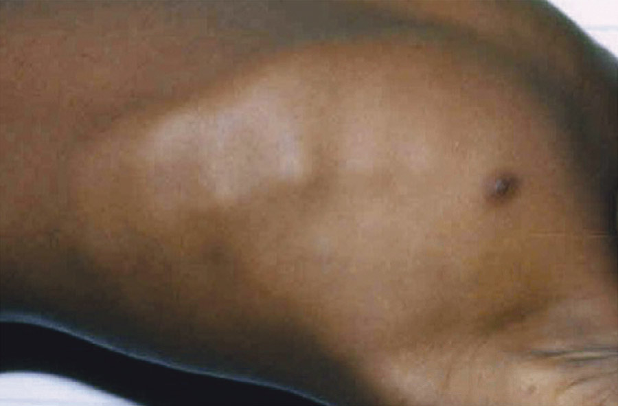
(Courtesy Dr. Thomas D. Thacher, Rochester, MN. In Kliegman RM et al: Nelson textbook of pediatrics, ed 22, Philadelphia, 2025, Elsevier.)
Figure E2 Hands and forearms of a young child with rickets.
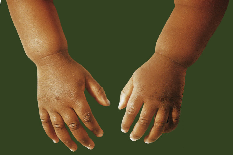
Prominence above the wrist is shown, resulting from flaring and poor mineralization of the lower end of the radius and ulna.
(From Bullough PG: In Kliegman RM et al: Nelson textbook of pediatrics, ed 22, Philadelphia, 2025, Elsevier.)
Figure E3 A, A Typical Example of Rickets
Note the Bowing of the Femurs and Tibiae, Which May Result from Vitamin D Deficiency, Phosphate Deficiency, or Other Causes. B, A Skeletal Radiograph of a Child with Rickets. Note that the Weight-Bearing Bones of the Lower Extremities are Bowed and that the Epiphyses are Open, Mottled, and Overgrown. C, Looser Zones or Pseudofractures that are Characteristic of Osteomalacia or Rickets. Because the Epiphyses are Closed, the Patient is an Adult. This Radiograph is Diagnostic of Osteomalacia.
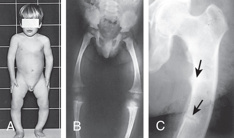
(From Stewart A: Metabolic bone diseases. In Andreoli TE et al [eds]: Cecil essentials of medicine, ed 8, Philadelphia, 2010, Saunders.)
TABLE E1 Clinical Features of Rickets
| General | |||
| Head | |||
| Chest | |||
| |||
| Back | |||
| Extremities | |||
| Hypocalcemic Symptoms† | |||
* These features are associated most frequently with vitamin D deficiency disorders.
† These symptoms develop only in children with disorders that produce hypocalcemia.
The less pronounced clinical presentation of adults with osteomalacia includes5:
The most common cause of rickets and osteomalacia is vitamin D deficiency. Insufficient calcium and phosphorus intake can also lead to rickets. Vitamin D and parathyroid hormone (PTH) play an important role in calcium homeostasis. Vitamin D is required for adequate calcium absorption in the gastrointestinal tract. Low vitamin D leads to reduced intestinal absorption of calcium, causing a compensatory increase in PTH and resorption of bone, leading to increased alkaline phosphatase levels.4 Vitamin D deficiency may arise from various conditions including inadequate dietary intake, malabsorption, chronic parenteral nutrition, and the additional risk factors listed previously. Malabsorption that causes inefficient mineralization is observed in diseases such as cystic fibrosis, intestinal disorders such as celiac disease, and in many chronic illnesses, including severe liver disease.4
Chronic renal failure can produce bone mineralization deficiency due to electrolyte abnormalities causing hypocalcemia and renal osteodystrophy (renal rickets). This results in decreased excretion of phosphate, therefore elevating serum phosphorus, along with elevated PTH and low levels of 1,25-OH vitamin D.1
Other causes specifically include vitamin D-dependent rickets (VDDR type I and II) and VDRR, also known as hereditary hypophosphatemic rickets5 (Table E2).
- •VDDR type I (VDDR-1) results from mutations in the vitamin D 1-alpha hydroxylase enzyme that converts inactive vitamin D to the active metabolite of 1,25(OH)2D.
- •VDDR type II (VDDR-II), also known as hereditary vitamin-D resistant rickets (HVDRR), results from defective vitamin D receptors due to a mutation in the vitamin D receptor gene (Table E3).
TABLE E3 Causes of Hypophosphatemic Rickets or Osteomalacia, or Both
| Genetic | |||
| Acquired | |||
From Melmed S et al: Williams textbook of endocrinology, ed 15, Philadelphia, 2025, Elsevier.
TABLE E2 Causes of Vitamin D-Deficiency Rickets or Osteomalacia, or Both
| Extrinsic | |||
| Intrinsic | |||
From Melmed S et al: Williams textbook of endocrinology, ed 15, Philadelphia, 2025, Elsevier.