section name header

Symptoms

Central scotoma, blurred or dim vision, objects may appear distorted (metamorphopsia) and miniaturized (micropsia), colors may appear faded. Usually unilateral, but can be bilateral (more likely in older patients, may not be symptomatic at the same time). May be asymptomatic.

Signs

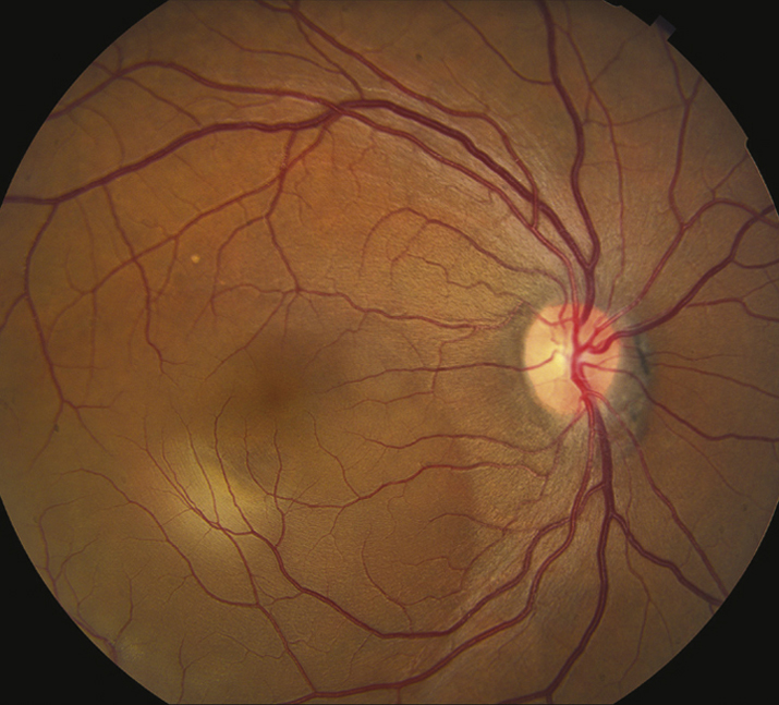
(See Figure 11.15.1.)

Critical

Localized serous detachment of the neurosensory retina in the macula without subretinal blood or lipid exudates. The margins of the detachment are sloping and merge gradually into the attached retina.

Other

Visual acuity usually ranges from 20/20 to 20/200. Amsler grid testing reveals relative scotoma and distortion of straight lines. May have a small RAPD, serous RPE detachment, or deposition of subretinal fibrin. Focal RPE changes may indicate sites of previous episodes.

11-15.1 Central serous chorioretinopathy.
Gervasio-ch011-image028

Differential Diagnosis

These entities may produce a serous detachment of the neurosensory retina in the macula

Etiology

  • Idiopathic: Most common in young adult to middle-aged men. In women, CSCR has an association with pregnancy. Increased incidence in patients with lupus.
  • Increased endogenous cortisol: This might explain a possible association with psychological or physiologic stress (type A personality). Rare cases exist with cortisol producing adrenal adenomas or Cushing syndrome.
  • Exogenous cortisol: Corticosteroid use, including nasal corticosteroid sprays and topical creams.

Work Up

Workup
  1. Amsler grid test to document the area of field involved. See Appendix 4, AMSLER GRID.
  2. Slit lamp examination of the macula with a fundus contact, Hruby, or 60- or 90-diopter lens to rule out concomitant CNV. In addition, search for an optic pit of the disc.
  3. Dilated fundus examination with indirect ophthalmoscopy to rule out a choroidal tumor or RRD.
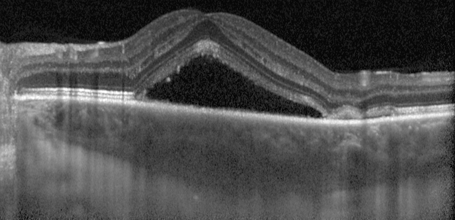
  4. OCT is helpful in demonstrating subretinal fluid or PEDs and for monitoring purposes. Enhanced-depth imaging OCT often demonstrates choroidal thickening and may be a useful adjunct in diagnosis (see Figures 11.15.2 and 11.15.3).
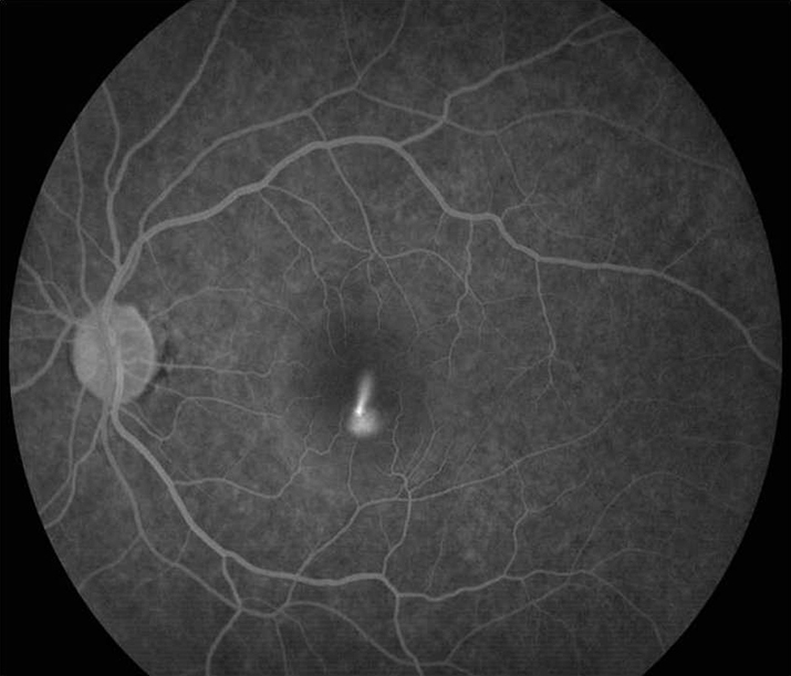
  5. IVFA and ICGA if the diagnosis is uncertain or presentation atypical, CNV is suspected, or laser treatment is to be considered. IVFA shows the nearly pathognomonic “smoke-stack” pattern of dye leakage in 10% to 20% of cases. ICGA shows choroidal artery and choriocapillaris filling delays and characteristic multifocal hyperfluorescent patches in the early phase.
  6. In cases of chronic CSCR, a systemic workup including cortisol levels and renal function should be considered.

11-15.3 Enhanced-depth imaging optical coherence tomography of central serous chorioretinopathy showing subretinal fluid and choroidal thickening.

Gervasio-ch011-image030

11-15.2 Intravenous fluorescein angiography of central serous chorioretinopathy showing “smoke-stack” pattern of dye leakage.

Gervasio-ch011-image029

Treatment

  1. Observation: Acute CSCR is usually self-limited with good visual prognosis. Worse prognosis for patients with recurrent disease, multiple areas of detachment, or prolonged course.
  2. Stop corticosteroids, if possible, including topical skin and nasal spray preparations.
  3. Laser photocoagulation: May accelerate visual recovery, but long-term benefit and safety are unclear. May increase risk of CNV formation. Given the CNV risk, use low laser intensity. Consider laser for:
    • Persistence of a serous detachment for several months.
    • Recurrence of the condition in an eye that sustained a permanent visual deficit from a previous episode.
    • Occurrence in the contralateral eye after a permanent visual deficit resulted from a previous episode.
    • Patient requires prompt restoration of vision (e.g., occupational necessity).
  4. Photodynamic therapy (PDT): May be helpful for chronic CSCR. Half-dose PDT may be considered for rapid SRF resolution in patients with acute CSCR.
  5. Mineralocorticoid receptor antagonists: Eplerenone and spironolactone have been associated with improved anatomic and visual outcomes in chronic CSCR.
  6. If CNV develops, consider anti-VEGF therapy.

Follow Up

  1. Examine patients every 6 to 8 weeks until resolution.