Pain, redness, and swelling over the lacrimal sac at the innermost aspect of the lower eyelid. Tearing, discharge, fever, or chills may also be present. Symptoms may be recurrent.
(see Figure 6.11.1.)
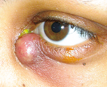
Erythematous, tender, tense swelling over the nasal aspect of the lower eyelid and extending around the periorbital area. Mucoid or purulent discharge may be expressed from the punctum when pressure is applied over the lacrimal sac, although this is not necessary nor sufficient for diagnosis.
Swelling in dacryocystitis is below the medial canthal tendon. Suspect lacrimal sac tumor (rare) if the mass is above the medial canthal tendon. |
Fistula formation may occur between the skin and the lacrimal sac. A lacrimal sac cyst or mucocele can occur in chronic cases. Progression to a lacrimal sac abscess, and rarely, orbital or facial cellulitis may occur.
Facial cellulitis involving the medial canthal area: The lacrimal drainage system is patent on irrigation. See 6.9, Preseptal Cellulitis.
Canaliculitis: Swelling and pain are limited to the canalicular and punctal region, typically does not extend over the lacrimal sac. Nasolacrimal duct is patent when lavaging through unaffected canaliculus. See 6.10, Canaliculitis.
Dacryocystocele: Mild enlargement of noninflamed lacrimal sac in an infant. Present at birth, but may not be detected until later. Caused by nasolacrimal duct obstruction or entrapment of mucus or amniotic fluid in the lacrimal sac and usually unilateral. If bilateral, assess breathing to rule out nasal obstruction. May also be acquired from chronic obstruction in adulthood. Conservative therapy with digital massage, antibiotic ointment, and warm compresses is usually sufficient for nonobstructive cases. Unless there is associated dacryocystitis, a dacryocystocele is nontender and nonerythematous.
Acute ethmoid sinusitis: Pain, tenderness, nasal obstruction, and erythema over the nasal bone, just medial to the inner canthus. Patients may be febrile. Imaging is diagnostic.
Frontal sinus mucocele/mucopyocele: Swelling typically occurs well above the medial canthal tendon. Proptosis, downward and lateral displacement of the globe, and external ophthalmoplegia are often present. Imaging is diagnostic.
Uncommon causes include lacrimal sac diverticula, dacryoliths, nasal or sinus surgery, trauma, and rarely lacrimal sac tumors.
Gram-positive bacteria (S. aureus and S. pneumoniae) are the most common pathogens; gram-negative and atypical organisms are seen more commonly in diabetics, immunocompromised, and nursing home patients.
History: Distinguish reflex tearing from epiphora. Previous episodes? Concomitant ear, nose, or throat infection? Underlying sinus disease? Prior trauma or surgery?
External examination: Apply gentle pressure to the lacrimal sac in the nasal corner of the lower eyelid with a cotton-tipped swab. Discharge, if present, may be expressed from the punctum. Perform bilaterally to uncover subtle contralateral dacryocystitis.
Evaluation for orbital signs: Assess pupillary response, extraocular motility, globe position for proptosis, and other evidence of potentially concurrent orbital cellulitis.
Obtain Gram stain and blood agar culture (consider chocolate agar culture in children given the higher incidence of Haemophilus influenzae) of any discharge expressed from the punctum or from incision and drainage of the lacrimal sac abscess.
Consider obtaining a complete blood count, lactate, and blood cultures in patients who appear acutely toxic.
Consider a CT scan of the orbits and paranasal sinuses in atypical cases, severe cases, suspected trauma, and those that do not respond to appropriate antibiotics.
Systemic antibiotics in the following regimen: Children older than 5 years and <40 kg:
Afebrile, systemically well, mild case, and reliable parent: Amoxicillin/clavulanate: 25 to 45 mg/kg/d p.o. in two divided doses for children, with a maximum daily dose of 90 mg/kg/d.
Alternative treatment: Cefpodoxime: 10 mg/kg/d p.o. in two divided doses for children, with a maximum daily dose of 400 mg.
Febrile, acutely ill, moderate-to-severe case, or unreliable parent: Hospitalize and treat with cefuroxime, 50 to 100 mg/kg/d i.v. in three divided doses in consultation with an infectious disease specialist.
Afebrile, systemically well, mild case, and reliable patient: Cephalexin 500 mg p.o. q6h or amoxicillin/clavulanate 500/125 mg t.i.d. or 875/125 mg p.o. b.i.d.
If exposure to MRSA is suspected, then start one to two tablets double-strength trimethoprimsulfamethoxazole 160/800 mg p.o. q12h for adults. Alternatively, start clindamycin 300 mg p.o. t.i.d. In addition to covering MRSA, this antibiotic also gives good coverage for anaerobes, streptococci, and methicillin-sensitive S. aureus.
Febrile, acutely ill, or unreliable: Hospitalize and treat with cefazolin 1 g i.v. q8h. See 7.3.1, Orbital Cellulitis.
The antibiotic regimen is adjusted according to the clinical response and culture/sensitivity test results. I.V. antibiotics can be changed to comparable p.o. antibiotics depending on the rate of improvement, but systemic antibiotic therapy should be continued for at least a full 10- to 14-day course.
Topical antibiotic drops (e.g., trimethoprim/polymyxin B q.i.d.) may be used in addition to systemic therapy. Topical therapy alone is not adequate.
Apply warm compresses and gentle massage to the inner canthal region for 5 to 10 minutes q.i.d.
Administer pain medication (e.g., acetaminophen with or without codeine) p.r.n.
Consider incision and drainage of a pointing abscess through the following steps:
The area of the lacrimal sac abscess is palpated and sterilized. Local 2% lidocaine with 1:100,000 epinephrine is infiltrated.
A curette is then used to break up any internal loculation and further pressure applied to ensure any further mucopurulence is expressed.
Small incision site is left open for further drainage. With ongoing drainage, a drain can be placed using iodoform gauze.
Once infection has resolved, evaluate patency of the nasolacrimal duct system with probing and irrigation. If an obstruction is present, consider surgical correction (e.g., dacryocystorhinostomy with silicone intubation). In cases of recurrent or chronic dacryocystitis, surgical correction is recommended.
Depends upon severity. If there is concern for progression to orbital cellulitis, then daily follow-up until improvement is confirmed may be needed. If outpatient condition worsens, hospitalization and i.v. antibiotics are recommended. Upon resolution of acute infection, probing and irrigation are required at the follow-up to assess the patency of the nasolacrimal drainage system.