Decreased vision, pain, tearing, foreign body sensation, photophobia, and redness. History of cataract surgery in the involved eye.
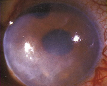
(See Figure 4.28.1.)
Corneal edema in an eye in which the native lens has been removed.
Corneal bullae, Descemet folds, subepithelial haze or scarring, corneal neovascularization, with or without preexisting guttae. Cystoid macular edema (CME) may be present.
Multifactorial: Corneal endothelial damage, intraocular inflammation, vitreous or subluxed intraocular lens or tube shunt touching (or intermittently touching) the cornea, preexisting endothelial dysfunction, and glaucoma.
Slit-lamp examination: Stain the cornea with fluorescein to check for denuded epithelium. Check the position of the intraocular lens if present, determine whether vitreous is touching the corneal endothelium, and evaluate the eye for inflammation. Check for subepithelial haze or scarring. Evaluate the fellow eye for endothelial dystrophy.
Consider a fluorescein angiogram or optical coherence tomography (OCT) to help detect CME, although evaluation may be limited by corneal opacification.
Topical sodium chloride 5% drops q.i.d. and ointment q.h.s., if epithelial edema is present.
Reduce IOP with medications if needed. Avoid epinephrine derivatives and prostaglandin analogs, if possible, because of the risk of CME (see 9.1, Primary Open-Angle Glaucoma).
Ruptured epithelial bullae (producing corneal epithelial defects) may be treated medically with the following:
Antibiotic drops (e.g., ofloxacin or moxifloxacin q.i.d.) or ointment (e.g., erythromycin or bacitracin q.i.d.) and a cycloplegic (e.g., cyclopentolate 1% t.i.d.). The topical antibiotic can be used even more frequently (e.g., q2h).
A bandage soft contact lens can be considered in conjunction with topical antibiotic drops (see 4.2, Recurrent Corneal Erosion).
Endothelial keratoplasty (DMEK or DSEK) or full-thickness corneal transplant (PK), possibly combined with intraocular lens repositioning, replacement, or removal and/or vitrectomy may be indicated when vision decreases or fails to recover, or when the disease becomes advanced and painful.
In patients with painful eyes and limited visual potential, anterior stromal micropuncture, excimer laser PTK, conjunctival flap, or amniotic membrane graft surgery may be considered.
See 11.14, Cystoid Macular Edema, for treatment of CME.