(see Figure 6.1.1.)
Concerning associated signs include anisocoria, proptosis, and ocular motility deficits. See individual entities.
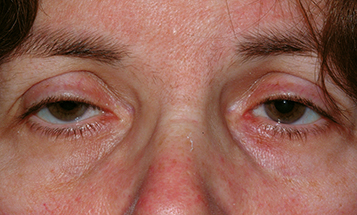
Drooping upper eyelid, superior visual field compromise. Peripheral visual compromise which often gets worse with reading and at night. Concerning associated symptoms include variability, diplopia, headache, and neck pain.
(see Figure 6.1.1.)
Concerning associated signs include anisocoria, proptosis, and ocular motility deficits. See individual entities.
Although the majority of ptosis is aponeurotic and of benign etiology, certain entities must be ruled out by careful examination. |
Third cranial nerve (CN) palsy (complete, partial, or aberrant CN III regeneration).
Chronic progressive external ophthalmoplegia (CPEO) (particularly Kearns–Sayre syndrome, see 10.12, Chronic Progressive External Ophthalmoplegia).
Categories
Myogenic: True myogenic ptosis is most commonly congenital. It is present at birth and is caused by localized dysgenesis of the levator palpebrae superioris. It results in poor levator function (<10 mm of excursion) and poor or absent eyelid crease. A poor Bell phenomenon (palpebral oculogyric reflex), lagophthalmos in downgaze, and upgaze limitation may indicate a double elevator palsy. Acquired myogenic ptosis is uncommon and may be seen with myotonic dystrophy, oculopharyngeal muscular dystrophy, and CPEO, or secondary to trauma.
Aponeurotic: The most common cause of ptosis. Occurs secondary to levator dehiscence and is characterized by an elevated eyelid crease, and preserved levator function (10 to 15 mm). Often worse in downgaze. Levator dehiscence is typically the result of involutional aging changes, but can be seen with repetitive eye rubbing, use of rigid contact lenses (pulling on the eyelids to put in or take out), trauma, or previous intraocular surgery (speculum-related muscle damage).
Neurogenic: Third CN palsy (often complete ptosis with pupillary and motility abnormalities; congenital, compressive, or vasculopathic, see 10.5, Isolated Third Nerve Palsy); Horner syndrome (mild, ∼2-mm upper and lower eyelid ptosis, see 10.2, Horner Syndrome); myasthenia gravis (variable degree, duration, and laterality, worsens with fatigue, see 10.11, Myasthenia Gravis); Marcus Gunn jaw-winking syndrome (ptotic eyelid elevates with jaw movement); ophthalmoplegic migraine; and multiple sclerosis.
Mechanical: Foreign-body or retained contact lens in upper fornix; upper eyelid or forniceal inflammation (chalazion, giant papillary conjunctivitis, posttraumatic or postsurgical edema); and neoplasm.
Traumatic: Eyelid laceration with levator involvement, levator contusion, tethering or ischemia within an orbital roof fracture, late dehiscence, or cicatricial changes.
Pseudoptosis: Contralateral eyelid retraction or proptosis, ipsilateral enophthalmos or hypertropia, microphthalmia, phthisis bulbi, dermatochalasis, brow ptosis, eyelid tumor, edema, blepharospasm, or Duane syndrome.
History: Onset and duration? Present since birth? Old photographs (e.g., driver’s license) and family members’ opinions are useful adjuncts to the history. Prior surgery in either eye? Trauma? Fluctuation throughout the day? Associated diplopia, headache, or neck pain? Trouble breathing or swallowing, associated drooling? History of autoimmune disease?
Mandatory documentation: Must carefully check and document pupillary size and extraocular motility, even if normal. If anisocoria is present, measurements should be documented under light and dark conditions. Additional pharmacologic testing may be indicated (see 10.1, Anisocoria). If extraocular muscle dysfunction is noted, additional testing with prism bars may be indicated.
Complete orbital examination: Measure and compare globe position with Hertel exophthalmometry, Proptosis may masquerade as contralateral ptosis. Resistance to retropulsion? Palpate superior orbit to rule out a mass or superior orbital rim deformity.
Complete eyelid examination: Margin reflex distance, levator function (full upper eyelid excursion with frontalis muscle held to inactivate brow recruitment), and upper eyelid crease position of both eyes. Is there lagophthalmos? Eyelid lag? Eyelid lag may masquerade as contralateral ptosis. Associated lower eyelid “reverse ptosis” (elevation of ipsilateral lower eyelid retractors) is often seen in Horner syndrome. Signs of aberrant eyelid movements such as jaw-winking, variability and/or fatigue, orbicularis weakness, and eyelid retraction with adduction and/or infraduction?
Complete ocular examination: Flip upper eyelid to examine conjunctival surface and superior fornix. Dilated fundus examination to look for pigmentary changes in adolescents and young adults who present with ptosis, poor levator function, and external ophthalmoplegia (possible CPEO and Kearns–Sayre syndrome).
Corneal protective mechanisms: Document presence or absence of lagophthalmos, orbicularis function, Bell phenomenon, and tear production. Check the cornea carefully for any abnormalities or dystrophies, which may predispose the patient to keratopathy.
Ice test: Apply ice pack to ptotic eye(s) for 2 minutes, measuring eyelid position before and after. Improvement in eyelid position is highly suggestive of myasthenia gravis.
Phenylephrine test: Instill one drop of 2.5% phenylephrine in the ptotic eye(s). Patients with an improvement of ptosis after 5 to 7 minutes may be good candidates for ptosis correction by Müller muscle-conjunctival resection.
Apraclonidine, cocaine, and hydroxyamphetamine tests. See 10.2, Horner Syndrome.
Imaging studies: In cases where a systemic or neurologic cause is suspected:
Computed tomography (CT) or magnetic resonance imaging (MRI) of orbit if an orbital process is suspected.
CT/computed tomography angiogram (CTA) or MRI/magnetic resonance angiogram (MRA) of the head and neck if Horner syndrome is present. This should be performed emergently if there is clinical suspicion for carotid artery dissection (neck pain, acute onset, and a history of trauma). Imaging of the head alone is inadequate. The extent of imaging must include the apex of the lung to adequately assess the entire sympathetic chain. See 10.2, Horner Syndrome.
Emergent CT/CTA, MRI/MRA, or conventional angiography to rule out posterior communicating artery aneurysm is indicated for all third CN palsies whether pupil-involving or pupil-sparing. See 10.5, Isolated Third Nerve Palsy.
CT chest if either Horner syndrome (to rule out apical lung mass compressing sympathetic ganglion) or myasthenia gravis (to rule out thymoma) is suspected. See 10.2, Horner Syndrome and 10.11, Myasthenia Gravis.
If myasthenia gravis is suspected, acetylcholine receptor antibody (binding, blocking, and modulating) testing, single-fiber electromyography (including the orbicularis muscle), and/or edrophonium chloride testing under monitored conditions may be indicated. See 10.11, Myasthenia Gravis.
Urgent ECG and cardiology consult if Kearns–Sayre syndrome is suspected. These patients can have heart block, resulting in sudden death.
Depends on the underlying etiology (see 10.2, Horner Syndrome; 10.5, Isolated Third Nerve Palsy; 10.11, Myasthenia Gravis).
Nonsurgical options: Observation. Oxymetazoline hydrochloride ophthalmic solution 0.1% can be applied topically to improve eyelid position (use with caution in patients at risk for angle-closure glaucoma). Taping upper eyelids open and eyelid crutches attached to glasses in neurogenic and myogenic ptosis.
Surgical options: External levator advancement, transconjunctival levator advancement, frontalis muscle suspension, Fasanella–Servat procedure, or Müller muscle-conjunctival resection (Müllerectomy). The surgical approach depends on preoperative evaluation and the underlying etiology of ptosis.
Congenital: Close follow-up is required to monitor for possible amblyopia (deprivation versus refractive secondary to induced corneal astigmatism), abnormal head positioning, and exposure keratopathy.
Traumatic: Observation for 6 months before considering surgical intervention. Many improve or completely resolve.
Chronic: Monitor for ptosis recurrence and exposure keratopathy.