section name header

Symptoms

Floaters, blurred vision, and/or flashes of light which are more common in dim illumination or with eye movement. Symptoms usually present acutely and can evolve over hours to days.

Signs

Critical

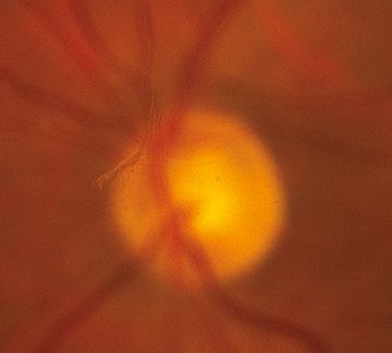
One or more discrete near-translucent or light gray vitreous opacities, one often in the shape of a ring (“Weiss ring”) or broken ring, suspended over the optic disc (See Figure 11.1.1).

Figure 11.1.1: Posterior vitreous detachment.

Rapuano9781975243722-ch011_f001.jpg

Other

Retinal break/tear (RT), retinal detachment (RD), pigmented cells in the anterior vitreous (“tobacco dust” or Shafer sign), or vitreous hemorrhage (VH) may occur with or without a posterior vitreous detachment (PVD), with similar symptoms. Peripheral retinal and disc margin hemorrhages.

NOTE

Approximately 10% of all patients with acute symptomatic PVD have a retinal break. The presence of pigmented cells in the anterior vitreous or VH in association with an acute PVD indicates a high probability (>70%) of a coexisting retinal break. See 11.2, Retinal Break (Tear).

Differential Diagnosis

Workup

  1. History: Duration of symptoms? Distinguish retinal photopsias from the visual phenomenon of migraine, which may be accompanied by new floaters. Presence and location of photopsias do not correlate with location of retinal break(s). Risk factors for retinal break (trauma, previous intraocular surgery, yttrium aluminum garnet [YAG] laser capsulotomy, high myopia, personal or family history of RT/RD)?

  2. Complete ocular examination, including evaluation of the vitreous for pigmented cells or VH and a dilated fundus examination with indirect ophthalmoscopy and scleral depression to rule out a retinal break and detachment. Optical coherence tomography (OCT) can be helpful in confirming the presence or absence of a PVD. Pseudophakic  patients may have smaller and more anterior breaks compared to phakic patients. Examine the fellow eye to assess for presence of PVD and peripheral retinal pathology. See Video: Scleral Depression Tutorial. See Video: Smartphone Video Indirect Ophthalmoscopy Guide.

  3. Visualize the PVD at the slit lamp with a handheld lens by identifying a gray-to-white ring or broken ring suspended in the vitreous. If not visible, have the patient make rapid saccades and then look straight to float the PVD into view.

  4. If VH obscures visualization of the retina, ultrasound (US) is indicated to identify the PVD and rule out a retinal break, RD, or other causes of VH. Inferior layering of VH may mimic a retinal break on US. See 11.13, Vitreous Hemorrhage.

Treatment

No treatment is indicated for PVD unless an acute retinal break or dense VH is found; see 11.2, Retinal Break (Tear).

NOTE

In the setting of acute PVD symptoms, consider treatment of chronic (pigmented) retinal breaks or lattice degeneration.

Follow-Up