Blurred vision, floaters, scotomas, metamorphopsia, and photopsias. Pain, redness, and photophobia are often absent due to lack of AC reaction.
Retinal or choroidal inflammatory lesions.
CME, ERM, choroidal neovascular membranes, peripheral neovascularization, retinal detachment, associated retinal vasculitis (sheathing and exudates).
Diagnostic Categories
CMV retinitis: Presents as a fulminant hemorrhagic necrosis, granular retinitis, or frosted-branch angiitis with minimal intraocular inflammation. See 12.12, Cytomegalovirus Retinitis.
Acute retinal necrosis (ARN). Most commonly secondary to VZV or HSV infection. Presents as large swaths of confluent retinitis that rapidly progresses and is typically associated with significant vitritis and anterior uveitis. See 12.13, Acute Retinal Necrosis.
PORN: Rapidly progressive herpetic outer retinal necrosis that occurs in immunocompromised patients, especially in advanced HIV/acquired immune deficiency syndrome (AIDS), with rapid involvement of the posterior pole and/or optic nerve over several days. Due to their immunocompromised status, it lacks significant signs of intraocular inflammation.
Bacterial endogenous endophthalmitis: Typically presents as a panuveitis, but more indolent bacteria can present with isolated posterior choroidal, subretinal, or retinal lesions alone. Lesions tend to be clustered in the posterior pole. See 12.17, Endogenous Bacterial Endophthalmitis.
TB: Most commonly bilateral multifocal, discrete, yellow lesions in the posterior pole. May be associated with optic nerve edema, vasculitis (classically a phlebitis, which may be occlusive). May present masquerading as tuberculosis serpiginous-like choroiditis.
Syphilis: Pleomorphic uveitis. Posterior placoid syphilis and punctate inner retinitis of syphilis are the more recognizable morphologies. See 12.10, Syphilis.
Candida endogenous endophthalmitis: Early discrete drusen-like choroidal lesions progressing to yellow-white, fluffy retinal, or preretinal lesions with a propensity for invading the vitreous cavity. See 12.18, Endogenous Fungal Endophthalmitis.
Aspergillus endogenous endophthalmitis: Early lesions resemble the feathered lesions of cotton-wool spots. Aspergillus has a propensity to invade the subretinal space.
Diffuse unilateral subacute neuroretinitis: Unilateral visual loss in children and young adults, caused by a subretinal nematode. Optic nerve swelling, vitreous cells, and deep gray-white retinal lesions are present initially but may be subtle. Later, optic atrophy, narrowing of retinal vessels, and atrophic RPE changes develop. Vision, visual fields, and electroretinogram deteriorate with time. Treatment is to laser the nematode. Antihelmintic drugs may be useful as adjunctive therapy.
Toxoplasmosis: Typically, unilateral retinal lesion which may or may not be associated with an adjacent pigmented chorioretinal scar. Associated with a dense vitritis over an area of white retinitis creating the classic headlight in a fog appearance See 12.9, Toxoplasmosis.
Sarcoidosis: Focal or multifocal initially creamy-appearing choroidal granulomas that progress to atrophic punched-out appearing lesions. May also be associated with optic nerve granulomas, retinal vasculitis (classically a phlebitis with candle-wax drippings) which may be occlusive and complicated by peripheral retinal neovascularization (see Figure 12.3.1).
White dot syndromes: See 12.8, White Dot Syndromes.
Autoimmune retinopathy: Subacute bilateral, but may be asymmetric; peripheral then central vision loss. OCT often shows progressive outer retinal loss. There is typically minimal vitritis or vasculitis. Late-stage findings include optic nerve pallor, macular edema, and retinal pigmentary changes.
Vitreoretinal lymphoma: Retinal findings include most commonly sub-RPE deposits that can be migratory in nature, leaving outer retinal and RPE atrophy in its wake. May be associated with vitreous cells.
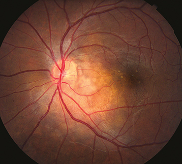
Neuroretinitis: Unilateral stellate macular exudates, optic nerve swelling, and vitreous cells. Classically caused by B. henselae (cat-scratch disease), but can also be caused by other etiologies including syphilis, sarcoidosis, Lyme disease, TB.
Obtain a thorough history and review of systems. In particular ask about a history of sexually transmitted diseases, high-risk sexual activity, intravenous drug use, fevers, chills, rash, difficulty breathing, travel, exposure to cats, neurologic symptoms (deficits or headaches), history of cancer or immunosuppression.
Complete ocular examination, including an IOP check, gonioscopy, and a dilated fundus examination.
Consider OCT to determine lesion location (intraretinal, subretinal, choroidal) and associated CME.
Consider an IVFA to detect subtle vasculitis, peripheral nonperfusion, peripheral neovascularization, or to monitor response to therapy.
Focused serologic testing based on history and examination:
Treponemal test (syphilis EIA, FTA-ABS, TP-PA), followed by confirmatory nontreponemal test (RPR, VDRL). See 12.10, Syphilis.
Interferon gamma releasing assay (IGRA, QuantiFERON Gold) and/or PPD. Consider chest imaging (CXR or CT chest) to assess for signs of active or prior pulmonary disease.
Serologic testing for antibodies against HSV, VZV, CMV, and toxoplasmosis can be useful in ruling out a disease as the causative etiology (IgG and/or IgM negative). The presence of positive IgG titer is not necessarily indicative of active disease, but rather prior exposure. Very elevated IgG titers may make you more suspicious for active disease, and a positive IgM indicates recent infection, but again are not fully diagnostic. Negative serologic testing can help rule out disease.
Consider blood cultures to detect bacteremia or fungemia; Urinalysis and urine cultures to detect a urinary tract infection (UTI).
Consider an AC paracentesis to detect DNA for CMV, HSV, VZV, or toxoplasmosis-associated disease. See Appendix 13, Anterior Chamber Paracentesis.
Consider MRI of the brain and orbits with and without gadolinium contrast to evaluate for comorbid CNS lesions if vitreoretinal lymphoma is of concern.
Consider referral to a vitreoretinal surgeon or ocular oncologist to perform a diagnostic vitrectomy ± retinal biopsy if concerned for possible vitreoretinal lymphoma. Specimens should be evaluated as soon as possible by an ocular pathologist or a neuropathologist with experience in working with vitreous specimens. Consider MYD88 mutation testing and IL-6/IL-10 ratio testing.
Consider topical prednisolone acetate 1% or difluprednate 0.05% q12h while awaiting workup results.
Treat infectious etiologies with appropriate antimicrobials prior to using local long-acting steroids or systemic steroids.
Oral acyclovir 400 mg five times per day or valacyclovir 500 mg t.i.d. for HSV-associated disease. Oral acyclovir 800 mg five times per day or valacyclovir 1000 mg t.i.d. for VZV-associated disease.
Oral valganciclovir 900 mg b.i.d. for CMV-associated disease.
For syphilis treatment, see 12.10, Syphilis.
For toxoplasmosis treatment, see 12.9, Toxoplasmosis.
For TB treatment, refer the patient to an internist, infectious disease specialist, or public health officer for consideration of systemic treatment. Patients with ocular TB frequently have no pulmonary disease but still require systemic four-drug antituberculous therapy. Concomitant oral steroids or steroid-sparing immunosuppression may be necessary.
Consider a trial of oral steroids after appropriate negative infectious workup.
Consider local steroid injection, particularly if the disease is unilateral, the eye is pseudophakic, and there is no history of steroid response ocular hypertension.
If the disease becomes persistent consider long-acting local steroids (0.19 mg or 0.59 mg fluocinolone acetonide implants) versus systemic steroid-sparing immunosuppression (e.g., antimetabolites, calcineurin inhibitors, and anti-TNF agents).
IVFA-guided panretinal photocoagulation should be considered in those with severe peripheral nonperfusion, active peripheral neovascularization, or complication from active peripheral neovascularization (e.g., vitreous hemorrhage).