section name header

Symptoms

Sudden, painless loss of vision or sudden appearance of black spots, cobwebs, or haze in the vision.

Signs

(See Figure 11.13.1.)

Figure 11.13.1: Vitreous and preretinal hemorrhage due to proliferative diabetic retinopathy.

Rapuano9781975243722-ch011_f024.jpg

Critical

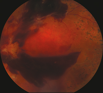
In severe VH, the red fundus reflex may be absent, and there may be limited or no view to the fundus. Red blood cells may be seen in the anterior vitreous (or anterior chamber). In mild VH, there may be a partially obscured view to the fundus. Chronic VH has a yellow-white appearance due to hemoglobin breakdown.

Other

A mild RAPD is possible in the setting of dense hemorrhage. Depending on the etiology, there may be other fundus abnormalities.

Differential Diagnosis

Etiology

NOTE

In infancy and childhood, consider birth trauma, child abuse (e.g., shaken baby syndrome), congenital X-linked retinoschisis, pars planitis, bleeding dyscrasias, and hematologic malignancies.

Workup

  1. History: Any ocular or systemic diseases? Trauma?

  2. Complete ocular examination, including slit-lamp examination with undilated pupils to check for iris neovascularization, IOP measurement, and dilated fundus examination of both eyes by using indirect ophthalmoscopy. In cases of spontaneous VH, scleral depression is performed if a retinal view can be obtained.

  3. When no retinal view can be obtained, B-scan US is performed to detect an associated RD or intraocular tumor. Flap RTs may be detected with scleral depression and may be seen on B-scan US (elevated flap). See Video: B-scan Ultrasound Tutorial.

  4. IVFA may aid in defining the etiology, although the quality of the angiogram depends on the density of the hemorrhage. Additionally, it may be useful to highlight abnormalities in the contralateral eye.

Treatment

  1. If the etiology of VH is not known and a retinal break or an RD or both cannot be ruled out (i.e., there is no known history of one  of the diseases mentioned previously, there are no changes in the contralateral eye, and the fundus is obscured by a total VH), close observation versus vitrectomy are options.

  2. Observation:

    • No heavy lifting, no straining, no bending. Keep head of bed elevated. This reduces the chance of recurrent bleeding and allows blood to settle inferiorly, permitting a view of the superior peripheral fundus, a common site for possible retinal breaks.

    • Discuss necessity of aspirin, nonsteroidal anti-inflammatory drugs (NSAIDs), and other anticlotting agents with prescribing physician. Consider stopping these agents when possible.

    • The underlying etiology is treated as soon as possible (e.g., retinal breaks are sealed with cryotherapy or laser photocoagulation, detached retinas are repaired, and proliferative retinal vascular diseases are treated with anti-VEGF therapy or laser photocoagulation).

  3. Vitrectomy:

    • VH accompanied by RD or RT on B-scan US.

    • Nonclearing VH. Because two-thirds of patients with an idiopathic, fundus-obscuring hemorrhage will have RTs or an RD, early vitrectomy should be considered.

    • VH with NVI.

    • Hemolytic or ghost cell glaucoma.

Follow-Up

If observation is elected, the patient is evaluated daily for the first 2 to 3 days. If a total, dense VH persists, and the etiology remains unknown, vitrectomy should be considered.