Infection of a filtering bleb. May occur any time after glaucoma filtering procedures (days to years). Greater incidence with use of antimetabolites during initial surgery, multiple surgeries, and postoperative complications including flat anterior chamber and wound leak.
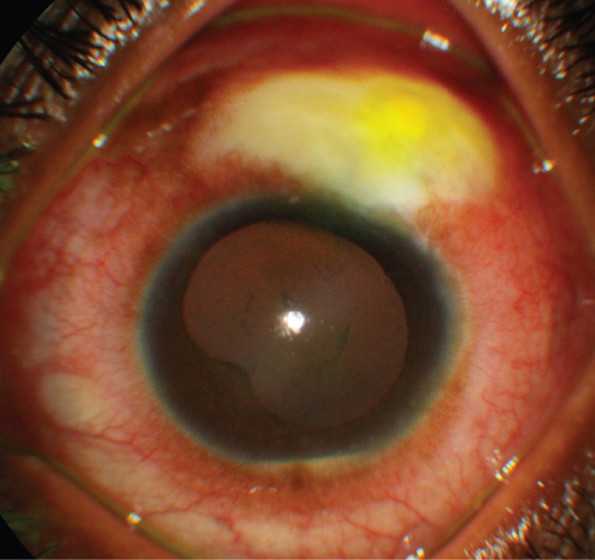
Grade 1 (mild): Bleb infection, hyperemia or purulence, but no anterior chamber or vitreous involvement.
Grade 2 (moderate): Bleb infection with anterior chamber inflammation but no vitreous involvement.
Grade 3 (severe): Bleb infection with anterior chamber and vitreous involvement. See 12.14, Acute Postoperative Endophthalmitis and12.15, Subacute and Chronic Postoperative Endophthalmitis.
Red eye and discharge early. Later, aching pain, photophobia, decreased vision, and mucous discharge.
(See Figure 9.18.1.)
Grade 1: Bleb appears milky with loss of translucency, microhypopyon in loculations of the bleb, may have frank purulent material in or leaking from the bleb, intense conjunctival injection. IOP is usually unaffected.
Grade 2: Grade 1 plus anterior chamber cell and flare, possibly an anterior chamber hypopyon, with no vitreous inflammation.
Grade 3: Grade 2 plus vitreous involvement. Same appearance as endophthalmitis except with bleb involvement.
Episcleritis: Sectoral inflammation, rarely superior. No bleb involvement. Minimal/mild pain. See 5.6, Episcleritis.
Conjunctivitis: Minimal decrease in vision, no pain or photophobia. Bacterial conjunctivitis can progress to blebitis if not promptly treated. See 5.1, Acute Conjunctivitis.
Anterior uveitis: Anterior chamber inflammation without bleb involvement. Photophobia. See 12.1, Anterior Uveitis (Iritis/Iridocyclitis).
Endophthalmitis: Similar findings as severe blebitis without bleb involvement. May have more intense pain, eyelid edema, chemosis, greater decrease in vision, and hypopyon. See 12.14, Acute Postoperative Endophthalmitis and 12.15, Subacute and Chronic Postoperative Endophthalmitis.
Ischemic bleb: Seen after the use of antimetabolites in immediate postoperative period. Conjunctiva is opaque with sectoral conjunctival injection.
Slit-lamp examination with careful evaluation of the bleb, anterior chamber, and vitreous. Search for bleb leak by performing a Seidel test (see Appendix 5, Seidel Test to Detect a Wound Leak). Look for microhypopyon with gonioscopy.
Culture bleb or perform anterior chamber tap for moderate blebitis. If severe, see 12.14, Acute Postoperative Endophthalmitis and 12.15, Subacute and Chronic Postoperative Endophthalmitis.
B-scan US will help identify vitritis if visualization is difficult.
Grade 1: Intensive topical antibiotics with:
Fortified cefazolin or vancomycin and fortified tobramycin or gentamicin every bour each alternating every half-hour for the first 24 hours. May begin with a loading dose of one drop of each, every 5 minutes and then repeated four times.
If fortified antibiotic drops are not available, fourth-generation (e.g., moxifloxacin, gatifloxacin, besifloxacin) fluoroquinolones q1h around the clock after a loading dose.
Consider use of oral fluoroquinolones as well (e.g., moxifloxacin 400 mg daily or ciprofloxacin 500 mg p.o. b.i.d.).
Reevaluate in 6 to 12 hours and again at 12 to 24 hours. Must not be getting worse.
May treat bleb leak with aqueous suppressants and cycloplegia.
Grade 2: Same approach as mild blebitis, plus cycloplegics, and more careful monitoring. May consider topical steroids after 24 to 48 hours of antibiotic therapy, pending improvement.
Grade 3: Treat as endophthalmitis with intravitreal tap and inject with some preference for early pars plana vitrectomy, as bleb-associated endophthalmitis appears to be more fulminant than infection following cataract surgery. See 12.14, Acute Postoperative Endophthalmitis and 12.15, Subacute and Chronic Postoperative Endophthalmitis.