section name header

Symptoms

Tenderness, redness, warmth, and swelling of the eyelid and periorbital area. Often there is a history of local skin abrasions, penetrating injury/trauma, hordeolum, or insect bites. Can be associated with sinusitis, although this is more common with postseptal infections. May complain of fever or chills.

Signs

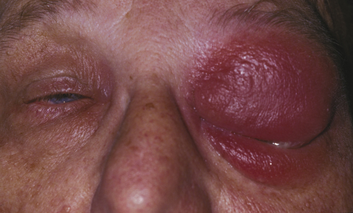
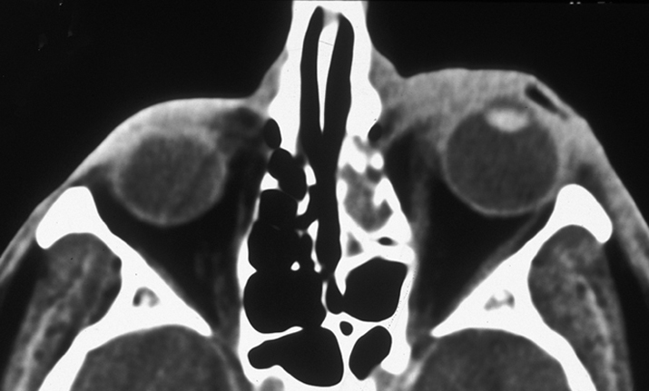
(See Figures 6.10.1 and 6.10.2.)

Critical

Eyelid erythema, tense edema, warmth, and tenderness. No proptosis, no optic neuropathy, no extraocular motility restriction, usually little to no conjunctival injection, and no pain with eye movement (unlike orbital cellulitis). The patient may not be able to open the eye because of eyelid edema. Visual changes such as blurred vision or monocular diplopia attributed to swollen eyelids.

Other

Tightness of eyelid skin or fluctuant eyelid edema. The eye itself may be slightly injected but is relatively uninvolved.

6-10.2 CT of preseptal cellulitis.
Gervasio-ch006-image008

6-10.1 Preseptal cellulitis.

Gervasio-ch006-image007

Differential Diagnosis

  • Orbital cellulitis: Proptosis, pain with eye movement, restricted motility, and possible visual compromise. See 7.3.1, ORBITAL CELLULITIS.
  • Chalazion: Focal inflammatory eyelid nodule. See 6.2, CHALAZION/HORDEOLUM.
  • Allergic: Sudden onset, nontender, itchy, and swollen eyelids. See 5.11, CONTACT DERMATITIS.
  • Erysipelas: Rapidly advancing streptococcal cellulitis, often with a clear demarcation line, high fever, and chills.
  • Necrotizing fasciitis: Severe bacterial infection involving subcutaneous soft tissue and deep fascia. Typically due to group A beta-hemolytic streptococci and S. aureus. Gray to purple skin discoloration with local hypesthesia is characteristic. Patients are often septic and may rapidly deteriorate. This potentially fatal condition requires emergent surgical debridement with i.v. antibiotic treatment.
  • Viral conjunctivitis with secondary eyelid swelling: Follicular conjunctivitis is present. See 5.1, ACUTE CONJUNCTIVITIS.
  • Cavernous sinus thrombosis: Proptosis with multiple cranial neuropathies. See 10.10, CAVERNOUS SINUS AND ASSOCIATED SYNDROMES (MULTIPLE OCULAR MOTOR NERVE PALSIES).
  • Varicella zoster virus: Vesicular rash that respects midline. See 4.16, HERPES ZOSTER OPHTHALMICUS/VARICELLA ZOSTER VIRUS.
  • Herpes simplex virus: Vesicular rash, typically unilateral, but does not respect midline/dermatomes. May be associated with ipsilateral follicular conjunctivitis. See 4.15, HERPES SIMPLEX VIRUS.
  • Other orbital disorders: Proptosis, globe displacement, or restricted ocular motility. See 7.1, ORBITAL DISEASE.
  • Others: Insect bite, angioedema, trauma, maxillary osteomyelitis, and others.

Etiology

  • Adjacent infection (e.g., hordeolum or dacryocystitis).
  • Trauma (e.g., puncture wound, laceration, insect bite).

Organisms

S. aureus and Streptococcus are most common, but H. influenzae should be considered in nonimmunized children. Suspect anaerobes if foul-smelling discharge or necrosis is present or if there is a history of an animal or human bite. Consider a viral cause if preseptal cellulitis is associated with a vesicular skin rash (e.g., herpes simplex or varicella zoster).

Work Up

Workup
  1. History: Pain with eye movements? Double vision? Prior trauma, sinus surgery or disease, cancer, chalazia, insect bites, or hair epilation? Sinus congestion or purulent nasal discharge? Recent vaccination? Prior antibiotic-resistant infections?
  2. Complete ocular examination: Evaluate for ocular motility restriction, proptosis, afferent pupillary defect, dyschromatopsia, and disc edema. An eyelid speculum or Desmarres eyelid retractor may facilitate ocular examination if the eyelids are excessively swollen.
  3. Check facial sensation in the distribution of first and second divisions of the trigeminal nerve.
  4. Palpate the periorbital area and the head and neck lymph nodes for a mass.
  5. Check vital signs.
  6. Obtain Gram stain and culture of any open wound or drainage.
  7. Perform CT scan of the orbits and sinuses (axial and coronal views) with contrast if there is a history of significant trauma or a concern for orbital or intraocular foreign body, orbital cellulitis, subperiosteal abscess, paranasal sinusitis, cavernous sinus thrombosis, or malignancy. Consider CT scan of the brain if any relevant history, signs, or symptoms necessitate this. Consider MRI or angiographic imaging if cavernous sinus or intracranial pathology is suspected, or if radiology recommends based on CT findings.
  8. Consider obtaining a complete blood count with differential to identify a leukocytosis and blood cultures in severe cases or when fever is present.

Treatment

  1. Antibiotic therapy
    1. Mild preseptal cellulitis, older than 5 years (<40 kg), afebrile, reliable patient/parent:
      • Amoxicillin/clavulanate: 25 to 45 mg/kg/d p.o. in two divided doses for children, a maximum daily dose of 90 mg/kg/d; 875/125 mg p.o. q12h for adults.
        or
      • Cefpodoxime: 10 mg/kg/d p.o. in two divided doses for children, a maximum daily dose of 400 mg; 200 mg p.o. q12h for adults.
        or
      • Cefdinir: 14 mg/mg/d p.o. in two divided doses for children with a maximum daily dose of 600 mg; 600 mg p.o. once daily for adults.
        If the patient is allergic to penicillin, then:
      • Trimethoprim/sulfamethoxazole: 8 to 12 mg/kg/d trimethoprim with 40 to 60 mg/kg/d sulfamethoxazole p.o. in two divided doses for children; 160 to 320 mg trimethoprim with 800 to 1600 mg sulfamethoxazole (one to two double-strength tablets) p.o. b.i.d. for adults.
        or
      • Moxifloxacin 400 mg p.o. daily (contraindicated in children). If exposure to MRSA is suspected, then:
      • Trimethoprim/sulfamethoxazole: 8 to 12 mg/kg/d trimethoprim with 40 to 60 mg/kg/d sulfamethoxazole p.o. in two divided doses for children; one to two tablets double-strength trimethoprim-sulfamethoxazole 160/800 mg p.o. q12h for adults.
        or
      • Doxycycline: 100 mg p.o. b.i.d. (contraindicated in children, pregnant women, and nursing mothers).
        or
      • Clindamycin: 10 to 30 mg/kg/d p.o. in three to four divided doses for children; 450 mg p.o. t.i.d. for adults. In addition to covering MRSA, this antibiotic also gives good coverage for streptococci and methicillin-sensitive S. aureus.
    2. Moderate-to-severe preseptal cellulitis or any one of the following:
      • Patient appears toxic.
      • Patient may be noncompliant with outpatient treatment and follow up.
      • Child 5 years or younger.
      • No noticeable improvement or worsening after 24 to 48 hours of oral antibiotics.
        Admit to the hospital for i.v. antibiotics as follows:
      • Vancomycin: 10 to 15 mg/kg i.v. every 6 hours in children; 0.5 to 1 g i.v. q12h for adults. (Dose adjustment is needed in cases of impaired renal function.)
        Plus one of the following:
      • Ampicillin/sulbactam: 300 mg/kg/d i.v. in four divided doses for children; 3.0 g i.v. q6h for adults.
      • Piperacillin–tazobactam: 240 mg/kg/d i.v. in three divided doses for children; 4.5 g i.v. q6h adults.
      • Ceftriaxone: 80 to 100 mg/kg/d i.v. in two divided doses for children with a maximum of 4 g/d; 2 g i.v. q12h for adults.
      • Cefepime: 50 mg/kg q12h (max 2 g/dose) in children; 1 to 2 g i.v. q12h in adults.
      • Ceftazidime: 90 to 150 mg/kg/day i.v. in three divided doses for children; 1 g i.v. q8–12h in adults.If the patient is allergic to penicillin, see 7.3.1, ORBITAL CELLULITIS, for alternatives.
  2. Warm compresses to the inflamed area t.i.d. p.r.n.
  3. Polymyxin B/bacitracin ointment to the eye q.i.d. if secondary conjunctivitis is present.
  4. Tetanus toxoid if needed. (See Appendix 2, TETANUS PROPHYLAXIS).
  5. Nasal decongestants if sinusitis is present.
  6. Orbital exploration and debridement are warranted if a fluctuant mass or abscess is present, or if a retained foreign body is suspected. Obtain Gram stain and culture of any drainage to guide antibiotic coverage. Avoid the orbital septum if possible. A drain may need to be placed.
  7. Consider an infectious disease consult for patients who have failed oral antibiotics and require i.v. treatment.
  8. Consider systemic steroids. As with orbital cellulitis, preseptal cellulitis will improve with proper antibiotic treatment of bacteria and surgical treatment of any nidus. In some cases, the inflammatory cascade caused by the infection is significant and may be slow to resolve. This may improve more quickly with a short course of steroids during or after antibiotic treatment for the infection. Steroid dosing and timing of initiation vary among clinicians. Physicians may start steroids simultaneously with antibiotics, after 24 to 48 hours of antibiotic coverage, or not at all.
NOTE:

Patients with the following risk factors should be covered for MRSA: history of MRSA infection or colonization, recurrent skin infections, contact with someone known to have MRSA, admission to a healthcare or long-term care facility within the past year, placement of a permanent indwelling catheter, on hemodialysis, i.v. drug use, incarceration within the past 12 months, participation in sports that include skin-to-skin contact or sharing of equipment and clothing, and poor personal hygiene.

NOTE:

Oral antibiotics are maintained for 10 to 14 days.

NOTE:

Intravenous antibiotics can be changed to comparable oral antibiotics after significant improvement is observed. Systemic antibiotics are maintained for a complete 10- to 14-day course. See 7.3.1, ORBITAL CELLULITIS, for alternative treatment. In complicated cases or patients with multiple allergies, consider consultation with an infectious disease specialist for antibiotic management.

Follow Up

Daily until clear and consistent improvement is demonstrated, then every 2 to 7 days until the condition has totally resolved. If preseptal cellulitis progresses despite antibiotic therapy, the patient is admitted to the hospital and a repeat (or initial) orbital CT scan is obtained. For patients already on p.o. antibiotics, switch to i.v. antibiotics (see 7.3.1, ORBITAL CELLULITIS).