section name header

Symptoms

Painless floaters and decreased vision. Minimal photophobia or external inflammation. Most often bilateral and classically affects patients aged 15 to 40 years.

Signs

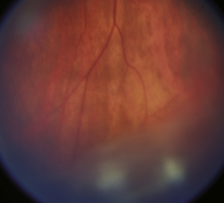
(See Figure 12.2.1.)

Critical

Vitreous cells and cellular aggregates floating predominantly in the inferior vitreous (snowballs). Younger patients may present with vitreous hemorrhage. White exudative material over the inferior ora serrata and pars plana (snowbank) is suggestive of pars planitis.

NOTE:

Snowbanking is typically in the inferior vitreous and can often be seen only with indirect ophthalmoscopy and scleral depression.

Other

Peripheral retinal vascular sheathing, peripheral neovascularization, mild anterior chamber inflammation, CME, posterior subcapsular cataract, band keratopathy, secondary glaucoma, ERM, and exudative retinal detachment. Posterior synechiae in pars planitis are uncommon and, if present, usually occur early in the course of the disease. Choroidal neovascularization is rare.

12-2.1 Pars planitis/intermediate uveitis with snowballs.
Gervasio-ch012-image002

Etiology

  • Pars planitis (>70%). Autoimmune disease of unknown cause in teenagers and young adults; insidious onset; bilateral but may be asymmetric; no clear gender predilection. CME most common cause of vision loss; vitreous hemorrhage may cause acute vision loss.
  • Sarcoidosis. See 12.6, SARCOIDOSIS.
  • Multiple sclerosis. See 10.14, OPTIC NEURITIS.
  • Lyme disease. See 13.3, LYME DISEASE.
  • Syphilis. See 12.12, SYPHILIS.
  • TINU: Rare cause of uveitis, mostly in young females but wide age distribution. Ocular disease usually follows renal disease but occasionally precedes or is concurrent. More commonly presents as anterior uveitis. Requires co-management with a nephrologist; eye disease may linger long after resolution of nephritis.
  • Primary intraocular lymphoma: intermediate uveitis may be the initial manifestation. Ask about neurologic symptoms in older patients; low threshold for CNS imaging. Referral to a retina specialist or an ocular oncologist for diagnostic pars plana vitrectomy and send the undiluted specimen to an experienced ocular cytopathologist; consider testing for interleukin-10 and MYD88 mutation. Oral steroids prior to biopsy may decrease the yield.
  • Toxocariasis. See 12.3, POSTERIOR UVEITIS and 8.1, LEUKOCORIA.
  • Others: IBD, Bartonellosis (cat-scratch disease), Whipple syndrome, primary Sjögren syndrome, etc.
  • Masquerade syndromes include retinoblastoma in children, old vitreous hemorrhage, and asteroid hyalosis.

Work Up

Workup
  1. Complete ocular examination. Includes gonioscopy to evaluate for possible neovascularization.
  2. Appropriate workup may include chest radiograph and/or chest CT, PPD and/or IGRA, ACE ± lysozyme, RPR or VDRL, and FTA-ABS or treponemal-specific assay. Obtain urinary beta-2 microglobulin, hemoglobin, creatinine, and urinalysis if TINU is suspected.
  3. Consider IVFA and/or OCT to document CME or retinal vasculitis.
  4. Consider lab testing for Lyme disease, toxoplasmosis, and cat-scratch disease in the appropriate clinical context. In older individuals, consider workup for malignancy/lymphoma.
  5. Consider magnetic resonance imaging (MRI) of the brain ± orbits with gadolinium to evaluate for demyelinating lesions if the review of systems is positive for current or previous focal neurologic deficits. Refer to a neurologist for multiple sclerosis workup if necessary.
Workup
  1. Complete history and review of systems: Ask about systemic disease or infection, skin rash, tattoos, intravenous drug abuse, indwelling catheter, risk factors for AIDS, recent eye trauma or surgery, travel (particularly to Ohio-Mississippi River Valley, Southwestern United States, New England, or Middle Atlantic area), and exposures (e.g., tick bite).
  2. Complete ocular examination, including IOP measurement and careful ophthalmoscopic examination. Indirect ophthalmoscopy with scleral depression of the ora serrata.
  3. Consider IVFA for diagnosis or therapeutic planning.
  4. Blood tests (any of the following may be obtained, depending on the suspected diagnosis, but a “shotgun” approach is inappropriate): Toxoplasma titer, ACE level, serum lysozyme, RPR or VDRL, FTA-ABS or treponemal-specific assay, ESR, ANA, ANCA, HLA-B51 (Behçet disease), HLA-A29 (birdshot retinochoroidopathy), Toxocara titer, IGRA ± PPD, and/or Lyme antibody. In neonates or immunocompromised patients, consider checking titers for HSV, VZV, CMV, and rubella virus. Cultures of blood and i.v. sites may be helpful when infectious etiologies are suspected. PCR techniques are available for HSV, VZV, CMV, and Toxoplasma.
  5. Chest radiograph or CT.
  6. CT/MRI of the brain and lumbar puncture when either lymphoma is suspected or there is any potential for central nervous system (CNS) involvement from an HIV-associated opportunistic infection.
  7. Diagnostic vitrectomy when appropriate (see individual sections).
  8. See individual sections for more specific guidelines on workup and treatment.

Treatment

Treat all vision-threatening complications (e.g., CME and vitritis) in symptomatic patients with active disease. Mild vitreous cell in the absence of symptoms, CME, or vision loss may be observed in cases of noninfectious intermediate uveitis.

  1. Topical prednisolone acetate 1% or difluprednate 0.05% q1–2h. Consider subtenon steroid (e.g., 0.5 to 1.0 mL injection of triamcinolone 40 mg/mL). May repeat the injections every 6 to 8 weeks until the vision and CME have stabilized. Slowly taper the frequency of injections. Subtenon steroid injections must be used with caution in patients with steroid-induced glaucoma. See Appendix 10, TECHNIQUE FOR RETROBULBAR/SUBTENON/SUBCONJUNCTIVAL INJECTIONS.
  2. If there is minimal improvement after three subtenon steroid injections 1 to 2 months apart, consider systemic steroids (e.g., prednisone 40 to 60 mg p.o. daily for 4 to 6 weeks), tapering gradually according to the patient’s response. High-dose systemic steroid therapy should last no longer than 2 to 3 months, followed by a taper to no more than 5 to 10 mg/d. Other options include sustained-release steroid implants (e.g., dexamethasone 0.7 mg intravitreal implant; fluocinolone acetonide 0.19 or 0.59 mg intravitreal implant) and immunomodulatory therapy (e.g., antimetabolites, calcineurin inhibitors, and anti-tumor necrosis factor agents), usually in conjunction with rheumatology.
  3. Transscleral cryotherapy to the area of snowbanking should be considered in patients who fail to respond to either oral or subtenon corticosteroids and who have neovascularization.
  4. Pars plana vitrectomy may be useful in cases refractory to systemic steroids or to treat vitreous opacification, tractional retinal detachment, ERM, and other complications. Additionally, vitreous biopsy through a pars plana vitrectomy may be indicated in cases of suspected masquerade syndromes, especially intraocular lymphoma.
NOTE:
  1. Some physicians delay steroid injections for several weeks to observe whether the IOP increases on topical steroids (steroid response). If a marked steroid response is found, depot injections should be avoided.
  2. Intravitreal preservative-free triamcinolone acetonide and the dexamethasone or fluocinolone acetonide implants are more effective for uveitic macular edema than periocular triamcinolone.
  3. Intravitreal anti-VEGF drugs (e.g., ranibizumab, bevacizumab, and aflibercept) are effective in the treatment of uveitic CME but only if the uveitis itself is suppressed.
  4. Oral acetazolamide 250 mg two to four times a day can reduce CME but is often not well tolerated due to nausea, diarrhea, fatigue, malaise, anorexia, or hypokalemia.
  5. Topical NSAIDs are usually not effective in patients with uveitic CME.
  6. Cataracts are a frequent complication of intermediate uveitis. If cataract extraction is performed, the patient should ideally be free of inflammation for 3 months preceding the operation. Consider starting the patient on oral prednisone 60 mg daily 2 to 5 days prior to surgery and tapering the prednisone over the next 1 to 4 weeks. Aggressive perioperative use of topical NSAIDS (e.g., ketorolac 0.5% q.i.d.) and topical steroids (e.g., prednisolone acetate 1% q2h or difluprednate 0.05% six times a day) starting 2 to 5 days preoperatively and continued for at least 4 to 6 weeks postoperatively may reduce the risk of pseudophakic CME and recurrent uveitis. Consider a combined pars plana vitrectomy at the time of cataract surgery if significant vitreous opacification is present.

Follow Up

  1. In the acute phase, patients are re-evaluated every 1 to 4 weeks, depending on the severity of the condition.
  2. In the chronic phase, re-examination is performed every 3 to 6 months. Monitor for neovascularization, CME, ERM, glaucoma, and cataract.

Other Causes of Vitreous Cells

  • Ocular ischemia.
  • Spillover from anterior uveitis.
  • Masquerade syndromes: Always consider these in the very old or very young patients.
    • Large cell lymphoma: Persistent vitreous cells in patients >50 years old, which usually do not respond completely to systemic steroids. Yellow-white subretinal infiltrates, retinal edema and hemorrhage, anterior chamber inflammation, or neurologic signs may be present.
    • Malignant melanoma: A retinal detachment and associated vitritis may obscure the underlying tumor. See 11.36, CHOROIDAL NEVUS AND MALIGNANT MELANOMA OF THE CHOROID.
    • Retinitis pigmentosa: Vitreous cells and macular edema may accompany waxy pallor of the optic disc, “bone-spicule” pigmentary changes, and attenuated retinal vessels. See 11.28, RETINITIS PIGMENTOSA AND INHERITED CHORIORETINAL DYSTROPHIES.
    • Rhegmatogenous retinal detachment (RRD): A small number of pigmented anterior vitreous and anterior chamber cells frequently accompany an RRD. Uveitis due to chronic retinal detachment (Schwartz–Matsuo syndrome) is one of the masquerade syndromes. See 11.3, RETINAL DETACHMENT.
    • Retained intraocular foreign body (IOFB): Persistent inflammation after a penetrating ocular injury. May have iris heterochromia. Diagnosed by indirect ophthalmoscopy, gonioscopy, B-scan ultrasonography (US), US biomicroscopy (UBM), or computed tomography (CT) scan of the globe. See 3.15, INTRAOCULAR FOREIGN BODY.
    • Retinoblastoma: Almost always occurs in young children. May also present with a pseudohypopyon and vitreous cells. One or more elevated white retinal lesions are usually, but not always, present. See 8.1, LEUKOCORIA.
    • Leukemia: Unilateral retinitis and vitritis may occur in patients already known to have leukemia. Prompt laboratory testing (complete blood count [CBC] with differential) and anterior chamber paracenteses (if there is anterior uveitis) for histology and immunostaining usually diagnostic.
    • Amyloidosis: Retrolenticular footplate-like deposits, vitreous globules, or vitreous membranes without any signs of anterior segment inflammation. Serum protein electrophoresis and diagnostic vitrectomy confirm the diagnosis. Rare.
    • Asteroid hyalosis: Small, white, refractile particles (calcium soaps) adherent to collagen fibers and floating in the vitreous. Usually asymptomatic and of no clinical significance.