section name header

Symptoms

Blurred vision and floaters. Scotomas and metamorphopsia are common. Photopsias are often present in posterior uveitis. Pain, redness, and photophobia are often present due to anterior chamber inflammation.

NOTE:

Panuveitis describes a pattern of severe, diffuse inflammation of both anterior and posterior segments. Often bilateral. Endophthalmitis or posterior scleritis should be considered in patients with posterior uveitis and significant pain.

Signs

Critical

Cells in the posterior vitreous, vitreous haze, retinal or choroidal inflammatory lesions, and retinal vasculitis (sheathing and exudates around vessels).

Other

Anterior and intermediate uveitis (indicative of panuveitis), retinal neovascularization, CME, ERM, and choroidal neovascular membranes.

Differential Diagnosis

Panuveitis

Possible etiologies are listed below:

White Dot Syndromes

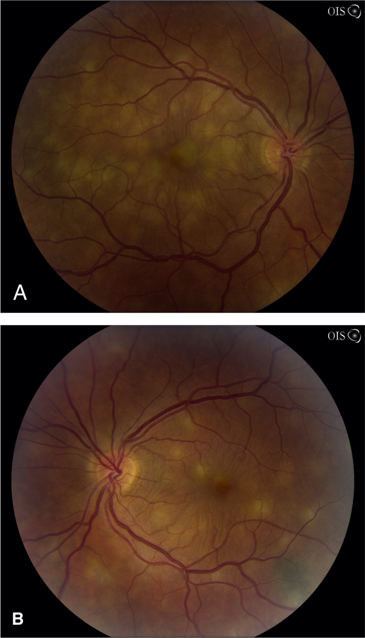
  • Acute posterior multifocal placoid pigment epitheliopathy (APMPPE): Acute visual loss in young adults, often after a viral illness. Multiple, creamy yellow–white or gray, plaque-like subretinal lesions in both eyes (see Figure 12.3.1A and B). Lesions block early and stain late on IVFA. Usually spontaneously improves over weeks to months without treatment. May be associated with a cerebral vasculitis (consider MRA if the patient has a headache or other neurologic symptoms), in which case, systemic steroids are indicated.
  • Multiple evanescent white dot syndrome (MEWDS): Photopsias and acute unilateral visual loss, often after a viral illness and usually in young women. May have a shimmering scotoma. Uncommonly bilateral, sequential, or recurrent. Characterized by multiple, small white lesions deep in the retina or at the level of the retinal pigment epithelium with foveal granularity and occasionally vitreous cells. Fluorescein angiography may show a classic perifoveal “wreath-like” pattern. There is often an enlarged blind spot on formal visual field testing. Vision typically returns to normal within 6 to 8 weeks without treatment.
  • Birdshot retinochoroidopathy: Usually middle-aged patients with multiple bilateral, oval, creamy-yellow spots deep to the retina, approximately 1 mm in diameter, scattered throughout the fundus but most prominent in inferior quadrants. A mild to moderate vitritis is present. Retinal vasculitis, CME, and optic nerve edema may be present. ICG angiography shows characteristic hypofluorescent spots but fluorescein angiography often shows only retinal vasculitis, CME, and “quenching” of dye. Positive HLA-A29 in 95% to 100% of patients. Early systemic immunosuppression is often recommended.
  • Multifocal choroiditis with panuveitis: Visual loss in young myopic women more often than men, typically bilateral. Multiple, small, round, and pale inflammatory lesions (similar to histoplasmosis) are located at the level of the pigment epithelium and choriocapillaris. Unlike histoplasmosis, vitritis occurs in 98% of patients. The lesions can occur in the macula and midperiphery and frequently respond to oral or periocular steroids, but typically recur with tapering, so that immunosuppressive therapy is often necessary. Choroidal neovascularization (CNV) occurs in about 1/3 of patients, and so patients should return for urgent evaluation if they have decreased vision or metamorphopsia.
  • Punctate inner choroidopathy: Blurred vision, paracentral scotoma, and/or photopsias, usually in young myopic women. Multiple, small round yellow-white spots predominantly in posterior pole with minimal intraocular inflammation. Lesions become well-demarcated atrophic scars within weeks. CNV may develop in up to 40% of patients. Systemic immunosuppression is usually indicated.
  • Serpiginous choroidopathy: Typically bilateral, recurrent chorioretinitis characterized by acute lesions (yellow-white subretinal patches with indistinct margins) bordering old atrophic scars. The chorioretinal changes usually extend from the optic disc outward; however, one-third may begin in the macula. Patients are typically aged 30 to 60 years. CNV may develop. Systemic immunosuppression is usually indicated. Must be distinguished from the “serpiginous” pattern of tuberculous chorioretinitis.
  • Toxocariasis: Typically unilateral. Usually occurs in children. The most common presentations are a macular granuloma (elevated white retinal/subretinal lesion) with poor vision, unilateral intermediate uveitis with peripheral granuloma, or endophthalmitis. A peripheral lesion may be associated with a fibrous band extending to the optic disc, sometimes resulting in macular vessel dragging. A severe vitritis and anterior uveitis may be present. A negative undiluted Toxocara titer in an immunocompetent host usually rules out this disease. See 8.1, LEUKOCORIA.
  • Presumed ocular histoplasmosis syndrome: Punched-out chorioretinal scars, peripapillary atrophy, CNV is common. Vitreous cells are absent. See 11.24, OCULAR HISTOPLASMOSIS.

12-3.1 Fundus photographs of right (A) and left (B) eye showing creamy yellow subretinal lesions in APMPPE.

Gervasio-ch012-image003

Note a pigmented choroidal nevus along the inferotemporal arcade in the left eye.

Retinitis

  • CMV retinitis: White patches of necrotic retina with granular borders, often mixed with retinal hemorrhage. Vascular sheathing (secondary frosted branch angiitis) in about 20% of eyes. Vitritis and anterior uveitis are usually mild. Seen in immunocompromised patients (most commonly in advanced HIV/AIDS, but also inherited or iatrogenic disorders of the immune system; rarely after periocular or intravitreal steroid injections) and congenitally infected neonates. See 12.9, CYTOMEGALOVIRUS RETINITIS.
  • Acute retinal necrosis (ARN): Unilateral or bilateral peripheral white patches of thickened necrotic retina with vascular sheathing that progress rapidly. Marked vitritis and anterior uveitis are typically present. See 12.8, ACUTE RETINAL NECROSIS.
  • Progressive outer retinal necrosis (PORN): Clinically similar to ARN, but may not have vitreous cells. Involves the posterior pole or optic nerve early, and classically spares the vessels. Occurs exclusively in severely immunocompromised patients, especially advanced HIV/AIDS, with rapid progression over several days. See 12.8, ACUTE RETINAL NECROSIS.
  • Toxoplasmosis: Unilateral retinal lesion may or may not be associated with an adjacent pigmented chorioretinal scar or clumps of scars. Focal dense vitritis over an area of white retinitis constitutes the “headlight in a fog” morphology. See 12.5, TOXOPLASMOSIS.
  • Candida: Early discrete drusen-like choroidal lesions progressing to yellow-white, fluffy retinal, or preretinal lesions. See 12.17, CANDIDA RETINITIS/UVEITIS/ENDOPHTHALMITIS.

Vasculitis

Retinal sheathing around vessels. Branch retinal vein and branch retinal artery occlusions may occur.

  • Periphlebitis (predominantly veins)
    • Sarcoidosis: Yellow “candlewax” exudates around veins.
    • Syphilis.
    • Pars planitis: Most prominent in the inferior periphery, neovascularization may be present.
    • Eales disease: Peripheral neovascularization and/or avascular retina.
    • Multiple sclerosis.
    • Birdshot retinochoroidopathy.
  • Arteritis (predominantly arteries)
    • Giant cell arteritis.
    • Polyarteritis nodosum.
    • Frosted branch angiitis.
    • Churg–Strauss disease.
    • ARN.
    • Idiopathic retinal vasculitis, aneurysms, and neuroretinitis (IRVAN).
    • Susac syndrome.
  • Both arteries and veins
    • Systemic lupus erythematosus.
    • Granulomatosis with polyangiitis (GPA, formerly Wegener granulomatosis).
    • Behçet disease.
    • HLA-B27-associated (rare).

Other Infectious Causes of Posterior Uveitis

  • Cat-scratch disease: Unilateral stellate macular exudates, optic nerve swelling, vitreous cells, and positive Bartonella serology. See 5.3, PARINAUD OCULOGLANDULAR CONJUNCTIVITIS.
  • Diffuse unilateral subacute neuroretinitis: Unilateral visual loss in children and young adults, caused by a nematode. Optic nerve swelling, vitreous cells, and deep gray-white retinal lesions are present initially, but may be subtle. Later, optic atrophy, narrowing of retinal vessels, and atrophic pigment epithelial changes develop. Vision, visual fields, and ERG deteriorate with time. Treatment is to laser nematode.
  • Lyme disease: Produces varied forms of posterior uveitis. See 13.3, LYME DISEASE.
  • Nocardia, Coccidioides species, Aspergillus species, Cryptococcus species, meningococcus, ophthalmomyiasis, onchocerciasis, and cysticercosis (seen more commonly in Africa and Central and South America).