section name header

Symptoms

  • Acute: Pain, redness, photophobia, consensual photophobia (pain in the affected eye when a light is shone in the fellow eye), excessive tearing, and decreased vision.
  • Chronic: Decreased vision (from cataract, vitreous debris, CME, or epiretinal membrane [ERM]) and floaters. May have periods of exacerbations and remissions with few acute symptoms (e.g., juvenile idiopathic arthritis [JIA]).

Signs

Critical

12-1.2 Grading of Anterior Chamber Flare

GradeDescription
0None
1+Faint
2+Moderate (iris/lens details clear)
3+Marked (iris/lens details hazy)
4+Intense (fibrin/plastic aqueous)

12-1.1 Grading of Anterior Chamber Cells

GradeCells in 1 × 1 mm Field
0<1
0.5+1 to 5
1+6 to 15
2+16 to 25
3+26 to 50
4+>50

Keratic Precipitates

  • Fine KP: Herpes simplex or varicella zoster virus, cytomegalovirus (CMV), Fuchs heterochromic iridocyclitis (FHIC).
  • Small, nongranulomatous KP (NGKP): HLA-B27-associated, trauma, masquerade syndromes, JIA, Posner–Schlossman syndrome (glaucomatocyclitic crisis), drug-induced. Granulomatous uveitides such as sarcoidosis can present with NGKP; the reverse rarely occurs.
  • Granulomatous KP (large, greasy, “mutton-fat”; mostly on the inferior cornea): Sarcoidosis, syphilis, tuberculosis (TB), JIA-associated, sympathetic ophthalmia, lens-induced, Vogt–Koyanagi–Harada (VKH) syndrome, and others.
  • CMV uveitis often has characteristic “coin-shaped” KP not found in other herpetic uveitides.
  • The location of KP can be helpful diagnostically.
    • Diffuse KP are characteristic of FHIC and herpetic uveitides.
    • KP underneath areas of stroma opacification suggest herpes simplex or, less often, varicella zoster keratouveitis.
    • Granulomatous KP in an inferior peripheral crescent underneath areas of stromal haze (often with fine, deep stromal vascularization) are highly suggestive of sarcoidosis.
    • Granulomatous KP in Arlt triangle (apex near central cornea, base at inferior limbus) are nonspecific.
    • “Crenated” KP are translucent and discrete, usually medium-large lesions characteristic of regressed granulomatous anterior uveitis.

Other

Low intraocular pressure (IOP) more commonly seen (secondary to ciliary body hyposecretion), elevated IOP can occur (e.g., herpetic, lens-induced, FHIC, Posner–Schlossman syndrome), fibrin (e.g., HLA-B27 or endophthalmitis), hypopyon (e.g., HLA-B27, Behçet disease, infectious endophthalmitis, rifabutin-induced, tumor), iris nodules (e.g., sarcoidosis, syphilis, TB), iris atrophy (e.g., herpetic, oral fourth-generation fluoroquinolones), iris heterochromia (e.g., FHIC), iris synechiae (especially HLA-B27, sarcoidosis), band keratopathy (especially JIA in younger patients, any chronic uveitis in older patients), uveitis in a “quiet eye” (consider JIA, FHIC, and masquerade syndromes), and CME (see Figure 12.1.1).
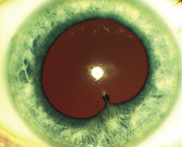
12-1.1 Anterior uveitis with posterior synechiae.

Gervasio-ch012-image001

Differential Diagnosis

  • Intermediate or panuveitis with spillover into the anterior chamber: Mainly floaters and decreased vision, positive fundoscopic findings (see 12.3, POSTERIOR UVEITIS).
  • Traumatic iritis. See 3.5, TRAUMATIC IRITIS.
  • Posner–Schlossman syndrome: Recurrent episodes of very high IOP and minimal inflammation. Many cases are caused by herpetic uveitis (herpes simplex virus [HSV], varicella zoster virus [VZV], and CMV). See 9.8, GLAUCOMATOCYCLITIC CRISIS/POSNER–SCHLOSSMAN SYNDROME.
  • Drug-induced uveitis (e.g., rifabutin, cidofovir, sulfonamides, pamidronate, systemic fluoroquinolones [especially moxifloxacin], biologic drugs, cancer immunotherapy [checkpoint inhibitors], and some chemotherapeutic drugs).
  • Sclerouveitis: Uveitis secondary to scleritis; typically presents with profound pain and tenderness to palpation. See 5.7, SCLERITIS.
  • Contact lens-associated red eye (CLARE): Red eye, corneal edema, epithelial defects, iritis with or without hypopyon, hypoxic subepithelial or stromal infiltrates (often multiple) may be present.
  • Infectious keratouveitis: Corneal infiltrate is present. See 4.11, BACTERIAL KERATITIS.
  • Infectious endophthalmitis: History of recent ocular surgery (including intravitreal injections), penetrating trauma, systemic infections (e.g., urinary tract infections), recent bowel or dental surgery, and skin wounds. Signs and symptoms include pain, hypopyon, fibrinous anterior chamber reaction, vitritis, decreased vision, and red eye; may have an endogenous source with fever, elevated white blood cell count. See 12.13 to 12.16, ENDOPHTHALMITIS SECTIONS.
  • Schwartz–Matsuo syndrome: Pigment released from a chronic retinal detachment clogs the trabecular meshwork, resulting in elevated IOP.
  • Tumor: Retinoblastoma and juvenile xanthogranuloma in children, primary intraocular lymphoma in elderly, metastatic disease in all ages, and others.
  • Pseudouveitis from pigment dispersion syndrome. Other findings include Krukenburg spindle and iris transillumination defects. Pigment cells in the anterior chamber are smaller than white blood cells and may disappear when viewed with a red-free light.

Etiology

  • Undifferentiated (idiopathic) (30% to 50% of anterior uveitis has no identifiable cause or disease association).
  • HLA-B27-associated uveitis: Systemic associations include ankylosing spondylitis, reactive arthritis (Reiter syndrome), psoriatic arthritis, and inflammatory bowel disease.
  • Lens-induced uveitis: Immune reaction to the lens material, often secondary to incomplete cataract extraction, trauma with lens capsule damage, or hypermature cataract. See 9.12, LENS-RELATED GLAUCOMA.
  • Postoperative iritis: Anterior chamber inflammation following intraocular surgery. Rule out acute endophthalmitis, retained lens fragments, iris chafing, or recurrence of preexisting anterior uveitis (e.g., HLA B27-associated uveitis). A small percentage of patients (especially African-Americans) with well-positioned posterior chamber intraocular lenses (IOL) may have a low-grade, steroid-responsive anterior uveitis that recurs when low-dose topical steroids are tapered off. Endophthalmitis must be considered if severe inflammation and pain are present. See 12.14, CHRONIC POSTOPERATIVE UVEITIS.
  • Uveitis–glaucoma–hyphema (UGH) syndrome: Usually secondary to irritation from an IOL (particularly a closed-loop anterior chamber lens or single-piece IOL in ciliary sulcus). A milder variant with iris chafing but no hyphema can occur (look for iris transillumination defects in an undilated pupil). See 9.16, POSTOPERATIVE GLAUCOMA.
  • Behçet disease: Young adults, acute simultaneous bilateral shifting hypopyon and iritis, aphthous ulcers, genital ulcerations, erythema nodosum, retinal vasculitis (arteries and/or veins), and hemorrhages, may have recurrent episodes.
  • VKH disease: acute unilateral or bilateral anterior uveitis occurs in chronic VKH disease.
  • Lyme disease: May have a history of a tick bite and rash. See 13.3, LYME DISEASE.
  • Anterior segment ischemia: Unilateral. Flare out of proportion to a cellular reaction. Corneal edema is common. Pain. Secondary to carotid insufficiency, tight scleral buckle, or previous extraocular muscle surgeries.
  • Tubulointerstitial nephritis and uveitis (TINU) syndrome: Uncommon but frequently underdiagnosed; usually bilateral nongranulomatous uveitis in children and young adults, female predilection. May be precipitated by oral nonsteroidal anti-inflammatory drug (NSAID) therapy. Systemic symptoms include abdominal pain, fatigue, and malaise. Urinary beta-2 microglobulin, urinary casts, and increased serum creatinine helpful diagnostically.
  • Toxoplasmosis: granulomatous unilateral anterior uveitis, posterior synechiae, and mutton-fat KP in Arlt triangle typically present. Occurs only with concurrent toxoplasmic retinitis.
  • Other rare infectious etiologies of anterior uveitis: Mumps, influenza, adenovirus, measles, chlamydia, leptospirosis, Kawasaki disease, rickettsial disease, chikungunya virus, and others. Ask about recent travel history.
NOTE:

Bilateral acute recurrent alternating anterior uveitis is very characteristic of HLA-B27 uveitis.

Chronic

  • JIA: Usually occurs in young girls with pauciarticular arthritis (4 joints involved); may be painless and asymptomatic with minimal injection. Usually bilateral. Iritis may precede typical arthritis. Positive antinuclear antibody (ANA), negative rheumatoid factor, and increased erythrocyte sedimentation rate (ESR) are most commonly seen. Associated with glaucoma, cataracts, band keratopathy, and CME. Uveitis less commonly occurs in polyarticular and rarely in systemic JIA (Still disease).
  • Chronic iridocyclitis of children: Usually occurs in young girls; it is similar to JIA in signs and symptoms but lacks arthritis.
  • FHIC: Floaters with or without blurred vision and glare, but few other symptoms, diffuse iris stromal atrophy often causing a lighter-colored iris with transillumination defects and blunting of the iris architecture. Gonioscopy may reveal fine vessels that cross the trabecular meshwork. Fine, stellate KP over the entire corneal endothelium, and mild anterior chamber reaction. Vitreous opacities, glaucoma, and cataracts are common, but macular edema and posterior synechiae are absent. Topical corticosteroids not helpful. Cataract surgery may cause anterior chamber hemorrhage from rupture of fine angle vessels, but outcomes are usually excellent.
  • Sarcoidosis: More common in African-Americans and Scandinavians. Usually bilateral; can have extensive posterior synechiae and conjunctival or iris nodules. See 12.6, SARCOIDOSIS.
  • HSV/VZV/CMV: May be chronic or acute and recurrent. Diffuse KP, increased IOP, and iris atrophy (transillumination defects). History of a unilateral recurrent red eye, occasionally history of skin vesicles, or history of shingles. Corneal scars associated with decreased corneal sensation may be present. HSV and VZV anterior uveitis usually require long-term oral acyclovir, valacyclovir, or famciclovir; CMV anterior uveitis usually requires oral valganciclovir. All three types usually require chronic low-dose topical corticosteroids for suppression.
  • Syphilis: Anterior and intermediate uveitis are most common. May have a maculopapular rash, iris roseola (vascular papules on the iris), and interstitial keratitis with ghost vessels in late stages. Inflammation of any ocular structure may occur. Placoid chorioretinitis is virtually pathognomonic. Neurosyphilis can cause meningismus. See 12.12, SYPHILIS.
  • TB: “Sticky” uveitis with extensive posterior synechiae, usually bilateral. Positive protein derivative of tuberculin (PPD) and/or interferon-gamma release assay (IGRA) (e.g., QuantiFERON-TB Gold), typical chest radiograph findings (helpful but not necessary for diagnosis; most TB uveitis occurs in patients without pulmonary TB), occasionally phlyctenular or interstitial keratitis, and sometimes signs of posterior uveitis. See 12.3, POSTERIOR UVEITIS.
  • Others: Leprosy, brucellosis, and other infectious causes.

Work Up

Workup
  1. Obtain a thorough history and review of systems. Specifically ask about fevers, chills, fatigue, malaise, cough, shortness of breath, joint pain/swelling/stiffness, diarrhea, blood in urine/stool, skin rashes, and oral or genital ulcers.
  2. Complete ocular examination, including an IOP check, gonioscopy, and a dilated fundus examination. The vitreous should be evaluated for cells.
  3. A laboratory workup may be unnecessary in certain situations:
    • First episode of a mild, unilateral, nongranulomatous uveitis with a history and examination that is not suggestive of systemic disease or herpetic uveitis.
    • Uveitis in the setting of known systemic disease such as sarcoidosis or the use of medicines known to cause uveitis (e.g., rifabutin).
    • Clinical findings are classic for a particular diagnosis (e.g., herpetic keratouveitis, FHIC, and toxoplasmosis).
  4. In all other situations requiring laboratory or diagnostic testing, a targeted workup is recommended. If too many tests are ordered unnecessarily, a portion of them may come back false-positive and confuse the diagnosis. See Table 12.1.3. However, if a patient presents with bilateral, granulomatous, or recurrent uveitis without a suspected diagnosis, our practice is to at least evaluate for sarcoidosis, syphilis, and TB (in at-risk patients). Consider additional workup as needed based on history and examination.
    • Syphilis testing (see 12.12 SYPHILIS).
    • PPD and/or IGRA. Limit use of TB testing to patients:
      • At risk of TB (e.g., immigrants from high-risk areas such as India, human immunodeficiency virus [HIV]-positive patients), homeless patients, or prisoners).
      • If immunosuppressive therapy (especially biologics) are being considered.
      • If anti-inflammatory therapy is not working.
    • Chest radiograph or chest CT to rule out sarcoidosis and pulmonary tuberculosis.
    • Angiotensin-converting enzyme (ACE) ± lysozyme (questionable utility).
    • Lyme antibody (consider in endemic areas).
    • HLA-B27 (in acute unilateral or bilateral alternating anterior uveitis; especially if hypopyon is present; ask about symptoms of a seronegative spondyloarthropathy).
    • Anterior chamber paracentesis for polymerase chain reaction (PCR) testing for suspected herpes virus-associated anterior uveitis (HSV, VZV, and CMV) and toxoplasmosis.
      • Serologic tests for infectious diseases have a low positive predictive value due to high seroprevalence in the general population, but negative tests (e.g., IgM and IgG for Toxoplasma gondii) may help to rule out the disease.

    12-1.3 Suggested Diagnostic Workup for Anterior Uveitis

    Ankylosing spondylitisHLA B27, SI joint films, rheumatology consult
    Reactive arthritisHLA B27, SI joint films (if symptomatic), swab for Chlamydia
    Psoriatic arthritisHLA B27, rheumatology and/or dermatology consult
    Lyme diseaseLyme antibody immunofluorescent assay (e.g., ELISA)
    Juvenile idiopathic arthritis or any suspect uveitis in childrenRheumatoid factor, antinuclear antibodies, HLA-B27, radiographs of affected joints, urinalysis and renal function tests, rheumatology consult
    SarcoidosisChest radiograph and/or chest CT, PPD or IGRA, ACE, lysozyme
    SyphilisRPR or VDRL, FTA-ABS or treponemal-specific assay; HIV testing if positive
    Ocular ischemic syndromeIntravenous fluorescein angiography, carotid Doppler studies
NOTE:

Autoimmune diseases are less common in the very young and very old—consider masquerades.

NOTE:

In children with uveitis, sarcoidosis and syphilis are much less common and no lab test should be ordered routinely except as indicated by the history and findings. Evaluation for systemic disease by a pediatric rheumatologist may be warranted (e.g., JIA and TINU).

Treatment

  1. Cycloplegic (e.g., cyclopentolate 1% t.i.d. for mild to moderate inflammation; atropine 1% b.i.d. to q.i.d. for severe inflammation).
  2. Topical steroid (e.g., prednisolone acetate 1%) q1–6h, depending on the severity of inflammation. Most cases of moderate to severe acute uveitis require q1–2h dosing initially. Difluprednate 0.05% may allow less frequent dosing than prednisolone acetate. Consider a loading dose (prednisolone acetate 1% one drop every minute for 5 minutes at bedtime and awakening) or fluorometholone 0.1% ophthalmic ointment at night. If the anterior uveitis is severe, unilateral, and is not responding to topical steroids, then consider periocular repository steroids (e.g., 0.5 to 1.0 mL subtenon injection of triamcinolone 40 mg/mL). See Appendix 10, TECHNIQUE FOR RETROBULBAR/SUBTENON/SUBCONJUNCTIVAL INJECTIONS.
  3. If there is no improvement on maximal topical and repository steroids, or if the uveitis is bilateral and severe, consider systemic steroids, or immunosuppressive therapy. Consider referral to a uveitis specialist and rheumatologist.
  4. Treat secondary glaucoma with aqueous suppressants. Avoid pilocarpine. Glaucoma may result from:
  5. If an exact etiology for the anterior uveitis is determined, then additional ocular and/or systemic management may be indicated.
    • Ankylosing spondylitis: Often requires systemic anti-inflammatory agents (e.g., NSAIDs such as naproxen). Consider consulting rheumatology, physical therapy, and cardiology (increased incidence of cardiomegaly, conduction defects, and aortic insufficiency).
    • Inflammatory bowel disease (IBD): Often benefits from systemic steroids, sulfadiazine, or other immunosuppressive agents. Obtain a medical or gastrointestinal consult.
    • Reactive arthritis (previously known as Reiter syndrome): If urethritis is present, then the patient and sexual partners are treated for chlamydia (e.g., single-dose azithromycin 1 g p.o.). Obtain medical and/or rheumatology or urology consult.
    • Psoriatic arthritis: Consider a rheumatology and/or dermatology consult.
    • Glaucomatocyclitic crisis: See 9.8, GLAUCOMATOCYCLITIC CRISIS/POSNER–SCHLOSSMAN SYNDROME.
    • Lens-induced uveitis: Usually requires removal of the lens material. See 9.12, LENS-RELATED GLAUCOMA.
    • Herpetic uveitis: Herpes simplex typically requires topical or oral antivirals and steroid drops for nonepithelial corneal disease. Herpetic iridocyclitis benefits from topical steroids and systemic antiviral medications (e.g., acyclovir, valacyclovir, or famciclovir); topical antivirals are usually ineffective for uveitis due to poor intraocular penetration. See 4.15, HERPES SIMPLEX VIRUS and 4.16, HERPES ZOSTER OPHTHALMICUS/VARICELLA ZOSTER VIRUS.
    • UGH syndrome: See 9.16, POSTOPERATIVE GLAUCOMA.
    • Behçet disease: See 12.7, BEHÇET DISEASE.
    • Lyme disease: See 13.3, LYME DISEASE.
    • JIA: Topical steroids can be useful acutely for reducing cells and flare, but should be minimized for long-term therapy to reduce the risk of cataract and glaucoma, both of which are more common in children. Systemic steroid therapy in children may cause growth suppression and should be avoided if possible. Prolonged cycloplegic therapy may be required and necessitate appropriate refractive correction. Consultation with rheumatology, pediatrics, and/or a uveitis specialist is useful as immunomodulatory therapy (e.g., methotrexate, adalimumab, and infliximab) is often needed. Regular follow up is essential, as flares may be asymptomatic; recurrent or chronic disease can lead to irreversible damage and various sequelae including synechiae, glaucoma (or hypotony), CME, epiretinal membrane, and cataract formation.
    • Chronic iridocyclitis of children: Same as JIA.
    • FHIC: Usually does not respond to or require steroids (a trial of steroids may be attempted, but they should be tapered quickly if there is no response); cycloplegics are not necessary.
    • Sarcoidosis: See 12.6, SARCOIDOSIS.
    • Syphilis: See 12.12, SYPHILIS.
    • Tuberculosis: Refer the patient to an internist, infectious disease specialist, or public health officer for consideration of systemic treatment. Patients with ocular TB frequently have no pulmonary disease but still require systemic four-drug antituberculous therapy. Concomitant oral steroids or methotrexate may be necessary.
NOTE:

The periocular use of triamcinolone is off-label and must be discussed with patients. A trial of topical steroids at full strength for several weeks may help identify patients at risk of a significant IOP increase from steroids. Additionally, periocular depot steroids should be used with extreme caution in patients with scleritis because of possible scleral melting.

NOTE:

Prior to giving periocular depot steroids, it is important to rule out infectious causes; oral steroids in such cases may be helpful starting 1 to 2 days after initiation of treatment for the underlying infection.

NOTE:

Cataract surgery in patients with JIA-associated uveitis has a high complication rate. Avoid cataract surgery if possible until the patient is inflammation-free for at least 3 months. An IOL may be placed in select circumstances and is preferable to aphakia in well-controlled disease.

NOTE:

Patients with FHIC usually do well with cataract surgery; however, they may develop a hyphema.

Follow Up

  1. Every 1 to 7 days in the acute phase, depending on the severity; every 1 to 3 months when stable.
  2. At each visit, the anterior chamber reaction and IOP should be evaluated.
  3. A vitreous and fundus examination should be performed for all flare-ups, when vision is affected, or every 3 to 6 months. Macular edema is a frequent cause of decreased vision even after the uveitis is controlled; OCT can be very useful.
  4. If the anterior chamber reaction has resolved, then the steroid drops can be slowly tapered with intermittent examinations to ensure that the inflammation does not return during the taper (usually one drop per day every 3 to 7 days [e.g., q.i.d. for 1 week, then t.i.d. for 1 week, then b.i.d. for 1 week, etc.]). Steroids are usually discontinued following the taper when the anterior chamber is quiet. Occasionally, long-term, low-dose steroids every day or every other day are required to keep the inflammation from recurring. Punctal occlusion techniques may increase the potency of the drug and decrease systemic absorption. The cycloplegic agents also can be tapered off as the anterior chamber reaction improves and no new posterior synechiae are noted.
NOTE:

Topical steroids should be tapered slowly to prevent severe rebound inflammation. If oral steroids are used, consider concurrent calcium 600 mg with vitamin D 400 units twice a day to reduce the risk of osteoporosis. In patients with very severe disease, note that doses of prednisone >60 mg/d increase the risk of ischemic necrosis of bone, and a three-day course of intravenous methylprednisolone 1 g/d for 3 days should be considered instead. Regular monitoring of glucose, blood pressure, lipids, and bone density should be done by a primary care doctor or rheumatologist if long-term oral steroid therapy is necessary.