section name header

Symptoms

Most are asymptomatic; can have decreased or distorted vision or both. Incidence increases with age. Twenty percent bilateral, though often asymmetric.

Signs

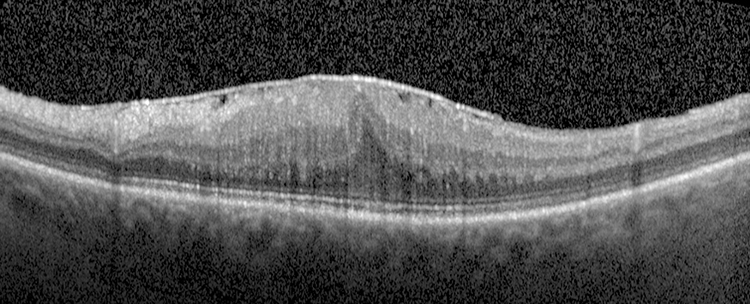
(See Figure 11.26.1.)

Figure 11.26.1: Epiretinal membrane with pseudohole.

Rapuano9781975243722-ch011_f047.jpg

Critical

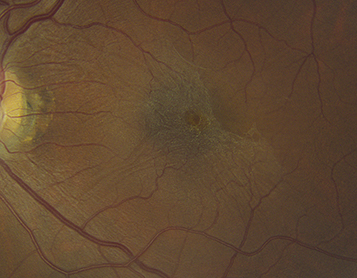
Spectrum ranges from a fine, glistening membrane (cellophane maculopathy) to a thick, gray-white membrane (macular pucker) present on the surface of the retina in the macular area.

Other

Retinal folds radiating out from the membrane; displacement, straightening, or tortuosity of the macular retinal vessels; ME or macular detachment. A ring-shaped condensation of the ERM around the fovea may simulate a macular hole (pseudohole).

Differential Diagnosis

Etiology

Workup

  1. History: Previous eye surgery or eye disease? Diabetes?

  2. Complete ocular examination, particularly a thorough dilated fundus evaluation and careful  macula evaluation with a slit lamp and a handheld lens. A careful peripheral examination should be performed to rule out a retinal break.

  3. OCT can show a hyperreflective layer over the surface of the retina, loss of foveal depression, and intraretinal cysts (See Figure 11.26.2).

    Figure 11.26.2: Optical coherence tomography of epiretinal membrane.

    Rapuano9781975243722-ch011_f048.jpg

Treatment

  1. Treat the underlying disorder.

  2. PPV with membrane peel can be considered when it significantly reduces vision.

Follow-Up

This is not an emergent condition, and treatment may be instituted at any time. Often does not progress over time. A small percentage of ERMs recur after surgical removal.