Binocular vertical (or oblique) diplopia, difficulty reading, sensation that objects appear tilted; may be asymptomatic.
(See Figures 10.7.1 and 10.7.2. See Video: Fourth Cranial Nerve Palsy.)
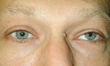
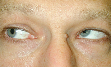
Figure 10.7.2: Isolated left fourth cranial nerve palsy: Right gaze with left inferior oblique overaction.

Deficient inferior movement of an eye when attempting to look down and in. The three-step test isolates a palsy of the superior oblique muscle (see #3 under Workup, Perform the three-step test).
The involved eye is higher (hypertropic) in primary gaze. The hypertropia increases when looking in the direction of the uninvolved eye or tilting the head toward the ipsilateral shoulder. The patient often maintains a head tilt toward the contralateral shoulder to eliminate diplopia.
All of the following may produce binocular vertical diplopia, hypertropia, or both:
Myasthenia gravis: Variable symptoms with fatigability. Ptosis common. Orbicularis oculi weakness often present. See 10.11, myasthenia gravis.
Thyroid eye disease: May have proptosis, eyelid lag, eyelid retraction, or injection over the involved rectus muscles. Positive forced duction test. See 7.2.1, Thyroid Eye Disease and Appendix 6, Forced Duction Test and Active Force Generation Test.
Idiopathic orbital inflammatory syndrome: Pain and proptosis are common. See 7.2.2, Idiopathic Orbital Inflammatory Syndrome.
Orbital fracture: History of trauma. Positive forced duction test. See 3.19, Orbital Blowout Fracture.
Skew deviation: The three-step test does not isolate a particular muscle. Rule out a posterior fossa or brainstem lesion with neuroimaging. See 10.13, Internuclear Ophthalmoplegia.
Incomplete third cranial nerve palsy: Inability to look down and in, usually with adduction weakness. Intorsion on attempted downgaze. Three-step test does not isolate the superior oblique. See 10.5, Isolated Third Cranial Nerve Palsy.
Brown syndrome: Limitation of elevation in adduction due to restriction of superior oblique tendon. May be congenital or acquired (e.g., trauma, inflammation). Positive forced duction test. See 8.8, Strabismus Syndromes.
GCA: Extraocular muscle ischemia causing nonspecific motility deficits or neural ischemia mimicking a cranial nerve palsy. Age ≥55 years, usually associated with systemic symptoms. See 10.17, Arteritic Ischemic Optic Neuropathy (Giant Cell Arteritis).
Trauma, vascular infarct (often the result of underlying diabetes or hypertension), congenital, or demyelinating disease.
History: Onset and duration of the diplopia? Misaligned eyes or head tilt since early childhood? Trauma? Stroke?
Examine old photographs to determine whether the head tilt is long standing, indicating a chronic or congenital fourth cranial nerve palsy.
Step 1: Determine which eye is deviated upward in primary gaze. This is best seen with the coveruncover test (see Appendix 3, Cover/Uncover and Alternate Cover Tests). The higher eye comes down after being uncovered.
Step 2: Determine whether the upward deviation is greater when the patient looks to the left or to the right.
Step 3: Determine whether the upward deviation is greater when tilting the head to the left shoulder or right shoulder.
Patients with a superior oblique muscle paresis have a hyperdeviation that is worse on contralateral gaze and when tilting the head toward the shoulder ipsilateral to the affected eye.
In addition to the findings on the three-step test, the hypertropia should be greater in downgaze than in upgaze.
Patients with bilateral fourth cranial nerve palsies demonstrate hypertropia of the right eye when looking left, hypertropia of the left eye when looking right, and a V-pattern esotropia (the eyes cross more when looking down due to a decrease of the abducting effect of the superior oblique muscles in depression as well as overaction of the inferior oblique muscles).
Perform the double Maddox rod test if bilateral fourth cranial nerve palsies are suspected to measure total excyclotorsion.
A white Maddox rod is placed before one eye and a red Maddox rod is placed before the other eye in a trial frame or phoropter, aligning the axes of each rod along the 90 degrees vertical mark. While looking at a white light in the distance, the patient is asked if both the white and red lines seen through the Maddox rods are horizontal and parallel to each other. If not, the patient is asked to rotate the Maddox rod(s) until they are parallel. If he or she rotates the top of this vertical axis outward (away from the nose) for more than 10 degrees total for the two eyes, then a bilateral superior oblique muscle paresis is likely present.
Measure vertical fusional amplitudes with a vertical prism bar to distinguish a congenital from an acquired palsy.
Ice test, rest test, or less commonly edrophonium chloride test if myasthenia gravis is suspected.
CT scan of head and orbits (axial, coronal, and parasagittal views) for suspected orbital disease.
Blood pressure measurement, fasting blood sugar, and hemoglobin A1c. Immediate ESR, CRP, and platelets if GCA is suspected. See 10.17, Arteritic Ischemic Optic Neuropathy (Giant Cell Arteritis).
An occlusion patch may be placed over one eye or fogging plastic tape can be applied to one lens of patients spectacles to relieve symptomatic double vision. Patching is usually not performed in children <11 years of age because of the risk of amblyopia.
Prisms in spectacles may be prescribed for small, stable hyperdeviations.
Strabismus surgery may be indicated for bothersome double vision in primary or reading position or for a cosmetically significant head tilt. Defer surgery for at least 6 months after onset of the palsy to allow for deviation stabilization or possible spontaneous resolution.
Acquired fourth cranial nerve palsy: As per the underlying disorder. If the workup is negative, the lesion is presumed vascular or idiopathic and the patient is reexamined in 1 to 3 months. If the palsy does not resolve in 3 months or if an additional neurologic abnormality develops, appropriate imaging studies of the brain are indicated. Patients are instructed to return immediately for any changes (e.g., ptosis, worsening diplopia, sensory abnormality, pupil abnormality).