section name header

Symptoms

Most often asymptomatic; can present with decreased or distorted vision, especially when CNV develops. Patients often have lived in or visited the Ohio–Mississippi River Valley or areas where histoplasmosis is endemic. Usually in the 20- to 50-year age range.

Signs

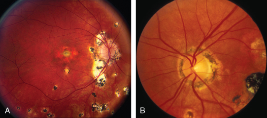
(See Figure 11.24.1.)

A, B: Ocular histoplasmosis.

Rapuano9781975243722-ch011_f043.jpg

Critical

Classic triad. Need two of the three to make the diagnosis:

  1. Yellow-white, punched-out round spots, chorioretinal scars, usually <1 mm in diameter  in any fundus location (histo-spots). Pigment clumps in or at the margin of the spots may be seen.

  2. A macular CNV appearing as a gray-green patch beneath the retina, associated with retinal edema, SRF, subretinal blood or exudate, or a pigment ring evolving into a disciform scar.

  3. Peripapillary atrophy or scarring, sometimes with nodules or hemorrhage. There may be a rim of pigment separating the disc from the area of atrophy or scarring.

Other

Curvilinear rows of small histo-spots in the peripheral fundus. No vitreous or aqueous cells.

Differential Diagnosis

Etiology

Fungal infection caused by Histoplasma capsulatum. Once acquired by inhalation, the organisms can pass to the choroid through the bloodstream. Importantly, ocular histoplasmosis is not thought to represent active infection and antifungal therapy is not indicated.

Workup

  1. History: Time spent in the Ohio–Mississippi River Valley or endemic area? Prior exposure to fowl?

  2. Amsler grid test (see Appendix 4, Amsler Grid) to evaluate the central visual field of each eye.

  3. Slit-lamp examination: Anterior chamber or vitreous cells and flare should not be present.

  4. Dilated fundus examination: Concentrate on the macular area with a slit lamp and a handheld lens. Look for signs of CNV and vitreous cells.

  5. IVFA and OCT to help detect CNV and monitor response to treatment.

Treatment

  1. Antifungal treatment is not helpful.

  2. Intravitreal anti-VEGF therapy for CNV is the mainstay of treatment. PDT for subfoveal CNV and focal laser photocoagulation for extrafoveal CNV may rarely be used as well.

Follow-Up

  1. Instruct all patients to use an Amsler grid daily and to return immediately if any sudden visual change is noted.

  2. Patients treated with anti-VEGF injections are seen every 4 to 6 weeks, depending on clinical response to therapy. Generally, a more complete treatment response is achieved with fewer injections than in AMD. Patients are often able to stop injections rather than undergoing lifelong treatment. Patients treated with PDT or focal laser are typically seen at 2 to 3 weeks, 4 to 6 weeks, 3 months, and 6 months after treatment, and then every 6 months thereafter.

  3. A careful macular examination and OCT is performed at each visit. IVFA may be repeated whenever renewed neovascular activity is suspected.

  4. Patients without CNV are seen every 6 months when macular changes are present in one or both eyes and yearly when no macular disease is present in either eye.