section name header

Symptoms

Decreased vision. Usually asymptomatic until middle adulthood.

Signs

(See Figure 11.22.1.)

Figure 11.22.1: High myopia with macular hemorrhage.

Rapuano9781975243722-ch011_f041.jpg

Critical

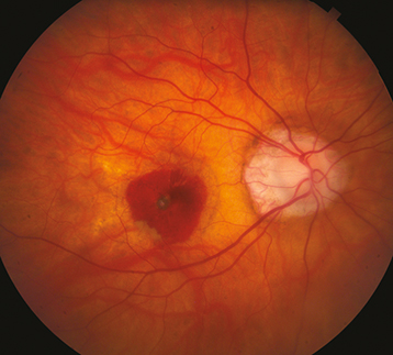
Myopic crescent (a crescent-shaped area of white sclera or choroidal vessels adjacent to the disc, separated from the normal-appearing fundus by a hyperpigmented line); an oblique (tilted) insertion of the optic disc, with or without vertical elongation. Macular pigmentary abnormalities; hyperpigmented spots in the macula (Fuchs  spots). Typically, a refractive correction of more than 6.00 diopters, axial length 26.5 mm.

Other

Increased risk for CNV or RRD. Temporal optic disc pallor, posterior staphyloma, entrance of the retinal vessels into the nasal part of the optic cup. The retina and choroid may be seen to extend over the nasal border of the optic disc. Well-circumscribed areas of atrophy, spots of subretinal hemorrhage, choroidal sclerosis, yellow subretinal streaks (lacquer cracks), peripheral retinal thinning, and lattice degeneration may occur. Visual field defects may be present.

Differential Diagnosis

Workup

  1. Manifest and/or cycloplegic refraction.

  2. IOP measurement by applanation tonometry (handheld tonometry may underestimate IOP in highly myopic eyes).

  3. Dilated fundus examination with indirect ophthalmoscopy to search for retinal breaks or detachment. Scleral depression may be helpful but should be performed with care over a staphyloma.

  4. Slit-lamp biomicroscopy with a handheld lens to examine macula and search for CNV (gray or green lesion beneath the retina, subretinal blood or exudate, or SRF).

  5. IVFA for suspected CNV.

  6. OCT can reveal CNV as well as macular detachment over a staphyloma. Additionally, OCT can be useful in identifying foveal schisis, a possible cause of vision loss in patients with high myopia.

Treatment

  1. Symptomatic retinal breaks are treated with laser photocoagulation, cryotherapy, or scleral buckling surgery. Treatment of asymptomatic retinal breaks may be considered when there is no surrounding pigmentation or demarcation line.

  2. Anti-VEGF agents are used for CNV due to high myopia. Laser photocoagulation therapy may be considered for extrafoveal or juxtafoveal CNV within several days of obtaining an IVFA, but is seldom performed. See 11.17,  Neovascular or Exudative (Wet) Age-Related Macular Degeneration.

  3. For glaucoma suspects, a single visual field often cannot distinguish myopic visual field loss from early glaucoma. Progression of visual field loss in the absence of progressive myopia, however, suggests the presence of glaucoma and the need for therapy. See 9.1, Primary Open-Angle Glaucoma.

  4. Recommend one-piece polycarbonate safety goggles for sports because of increased risk of choroidal rupture from minor trauma.

Follow-Up

In the absence of complications, re-examine every 6 to 12 months, watching for the related disorders discussed above.