AUTHOR: David J. Lucier Jr., MD, MBA, MPH
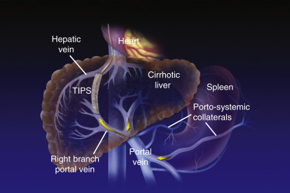
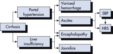
Cirrhosis is defined histologically as the presence of irreversible fibrosis in the liver. It can be classified as micronodular, macronodular, or mixed; however, each form may be seen in the same patient at different stages of the disease, and this classification system has little utility in determining the underlying etiology of cirrhosis. Cirrhosis manifests clinically with portal hypertension, ascites and peripheral edema, hepatic encephalopathy, and variceal bleeding.
|
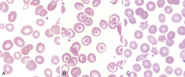
Jaundice, palmar erythema, spider angiomata, ecchymosis (thrombocytopenia or coagulation factor deficiency), increased pigmentation (hemochromatosis), xanthomas (primary biliary cirrhosis), and diffuse pruritus. Cutaneous lesions often accompany cirrhosis and can be found in >40% of people with chronic alcoholism
Kayser-Fleischer rings (corneal copper deposition seen in Wilson disease; best diagnosed with slit lamp examination), scleral icterus
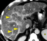
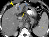
Tender or nontender hepatomegaly (congestive hepatomegaly), small, nodular liver (cirrhosis), palpable, nontender gallbladder (neoplastic extrahepatic biliary obstruction), palpable spleen (portal hypertension), dilated superficial periumbilical vein (caput medusae), venous hum auscultated over periumbilical veins (portal hypertension), ascites (portal hypertension, hypoalbuminemia), diffuse abdominal tenderness (spontaneous bacterial peritonitis)
Hemorrhoids (portal hypertension), guaiac-positive stools (alcoholic gastritis, bleeding esophageal varices, peptic ulcer disease, bleeding hemorrhoids, portal gastropathy)
In addition to an assessment of liver function, the evaluation of patients with cirrhosis should also include an assessment of renal and circulatory function. Diagnostic workup is aimed primarily at identifying the most likely cause of cirrhosis. The history is extremely important:
TABLE 1 Hemostatic Balance in Liver Disease
Promotes Thrombosis | Promotes Bleeding | |
---|---|---|
Primary hemostasis | ||
Secondary hemostasis | ||
Fibrinolysis |
ADAMST13, A disintegrin and metalloproteinase with thrombospondin; PAI-1, plasminogen activator inhibitor-1; TAFI, thrombin activatable fibrinolysis inhibitor; t-PA, tissue plasminogen activator; vWF, von Willebrand factor.
From Hoffman R et al: Hematology: basic principles and practice, ed 7, Philadelphia, 2018, Elsevier.
TABLE 2 Hemostatic Indices in Liver Disease
Laboratory Changes | PT | PTT | TCT | Fib | Clauss | Plt | Platelet Aggregation | FVII | DD | ELT |
---|---|---|---|---|---|---|---|---|---|---|
Thrombocytopenia | N | N | N | N | N | ↓ | N | N | N | N |
Platelet dysfunction | N | N | N | N | N | N | Abnormal | N | N | N |
Vitamin K deficiencya | ↑ | ↑ | N | N | N | N | N | ↓ | N | N |
Factor deficiency | ↑ | ↑ | N | N | N | N | N | ↓ | N | N |
Hypofibrinogenemia | N/↑ | N/↑ | ↑ | ↓ | ↓ | N | N | N | N | N |
Dysfibrinogenemia | N/↑ | N/↑ | ↑ | N | ↓ | N | N | N | N | N |
Hyperfibrinolysis | N/↑ | N/↑ | N/↑ | N/↓ | N/↓ | N | N | ↓ | ↑ | ↓ |
DIC | N/↑ | N/↑ | N/↑ | ↓ | ↓ | ↓ | N | N/↓ | ↑ | ↓ |
Clauss, Clauss fibrinogen; DD, D-dimer; DIC, disseminated intravascular coagulation; ELT, euglobulin lysis time (measure of fibrinolysis); Fib, fibrinogen; FVII, factor VII functional assay; N, normal; Plt, platelet; PT, prothrombin time; PTT, partial thromboplastin time; TCT, thrombin clotting time.
a Differentiating between vitamin K deficiency and liver disease can be challenging with conventional laboratory tests. If available, performing a factor II assay with and without Echis venom (factor II biologic and factor II Echis) may be useful. Ecarin is derived from Echis carinatus snake venom and can activate prothrombin irrespective of γ-carboxylation. Factor II activity (biologic) is reduced in both vitamin K deficiency and liver disease. In contrast, the factor II Echis is reduced in liver disease but is normal in vitamin K deficiency.
From Hoffman R et al: Hematology: basic principles and practice, ed 7, Philadelphia, 2018, Elsevier.
A, Macrocytes have a Mean Corpuscular Volume Greater than 100 Fl and Can Be Oval-Shaped. Commonly Associated Disorders Include Liver Disease and Vitamin B12 and Folate Deficiency. B, Target Cells are Characterized by the Bulls-Eye Appearance of the Red Blood Cell (RBC). They are a Result of an Increased Surface-to-Volume Ratio Related to Excess RBC Membrane (E.g., Liver Disease) or Disproportionate Reduction of Cytoplasmic Content (E.g., Hemoglobinopathies, Iron Deficiency). C, Acanthocytes, or Spur Cells, Typically Appear as Contracted RBCs Lacking Central Pallor with Multiple Irregular Membrane Projections. The Morphologic Appearance Reflects the Irreversible Cytoskeletal Damage that Has Occurred Because of Passage of Nondeformable RBCs through the Reticuloendothelial System. They are Most Commonly Seen in Severe Liver Disease but Can Also Be Features of Rare Neuroacanthocytosis Syndromes or Lipoprotein Disorders.
From Hoffman R et al: Hematology: basic principles and practice, ed 7, Philadelphia, 2018, Elsevier.
Delayed Portal Venous-Phase Computed Tomography Reveals that the Liver is Misshapen and Nodular in Contour. Parenchymal Density that is Significantly Lower than that of the Spleen (S) is Indicative of Fatty Infiltration and Continuing Liver Injury. Prominent Scars and Bands of Fibrosis (Arrowheads) are Seen Throughout the Liver. Ascites (a) is Present. (From Webb Wr Et Al: Fundamentals of Body CT, Ed 4, Philadelphia, 2015, Saunders.)
Postcontrast Computed Tomography Reveals a Liver that is Nodular in Contour (Arrowhead) with Patent Enlarged Paraumbilical Veins (Blue Arrows) and Splenomegaly (S), Findings Indicative of Portal Hypertension. Mildly Enlarged Portosystemic Collateral Vessels (Curved Arrow) are Also Evident in the Gastrohepatic Ligament.
From Webb WR et al: Fundamentals of body CT, ed 4, Philadelphia, 2015, Saunders.
This Shunt is Performed by Interventional Radiologists and Consists of an Expandable Metal Stent (Most Often Coated with Polytetrafluoroethylene) that Connects a Branch of the Portal Vein (High-Pressure Vein) to a Branch of the Hepatic Vein (Low-Pressure Vein). The Shunt Decompresses the Portal System and Portosystemic Collaterals, and Therefore It is Used in the Treatment of Selected Patients with Cirrhosis and Variceal Hemorrhage. The Shunt Also Decompresses the Hepatic Sinusoids and Therefore is Also Used in the Treatment of Refractory Ascites.
From Goldman L, Schafer AI: Goldman-Cecil medicine, ed 26, Philadelphia, 2019, Elsevier.
BOX 2 Management of Coagulopathy in Liver Disease
From Hoffman R et al: Hematology: basic principles and practice, ed 7, Philadelphia, 2018, Elsevier.
Afp, -Fetoprotein; Bm, Bowel Movement; D/C, Discontinue; Egd, Esophagogastroduodenoscopy; GI, Gastrointestinal; HCC, Hepatocellular Carcinoma; INR, International Normalized Ratio; Na, Sodium; NSAIDs, Nonsteroidal Antiinflammatory Drugs; R/O, Rule Out; Sbp, Spontaneous Bacterial Peritonitis; Us, Ultrasound.
From Goldman L, Schafer AI: Goldman-Cecil medicine, ed 26, Philadelphia, 2019, Elsevier.
Varices and Variceal Hemorrhage are a Direct Consequence of Portal Hypertension. Ascites Results from Sinusoidal Portal Hypertension and Can Be Complicated by Infection (Spontaneous Bacterial Peritonitis [sbp]) or Renal Dysfunction (Hepatorenal Syndrome [HRS]). Hepatic Encephalopathy Results from Portosystemic Shunting (I.e., Portal Hypertension) and Liver Insufficiency. Jaundice Results Solely from Liver Insufficiency.
From Goldman L, Schafer AI: Goldman-Cecil medicine, ed 26, Philadelphia, 2019, Elsevier.
BOX E1 Causes of Hepatic Decompensation in Cirrhosis
UTI, SBP, pneumonia, primary bacteremia, cellulitis Renal failure, hyponatremia, uremia, volume depletion (variceal bleed) Alcohol, acetaminophen (paracetamol) especially Also NSAIDs, diuretics, benzodiazepines, opiates, phenothiazines, anticonvulsants Hepatitis A, B, C, D; cytomegalovirus, Epstein-Barr virus |
From Talley NJ et al: Essentials of internal medicine, ed 4, Chatswood, NSW, 2021, Elsevier Australia.
TABLE 3 The Two Most Commonly Used Scoring Systems in Cirrhosis
1. Child-Turcotte-Pugh (CTP) Score (Range, 5-15) | |||
---|---|---|---|
Points Ascribed | |||
Parameters | 1 | 2 | 3 |
Ascites | None | Grade 1-2 (or easy to treat) | Grade 3-4 (or refractory) |
Hepatic encephalopathy | None | Grade 1-2 (or induced by a precipitant) | Grade 3-4 (or spontaneous) |
Bilirubin (mg/dl) | <2 | 2-3 | >3 |
Albumin (g/dl) | >3.5 | 2.8-3.5 | <2.8 |
Prothrombin time (seconds >control) or INR | <4 <1.7 | 4-6 1.7-2.3 | >6 >2.3 |
CTP classification: Child A: Score of 5-6; Child B: Score of 7-9; Child C: Score of 10-15 | |||
2. Model of End-Stage Liver Disease (MELD) Score (Range, 6-40) | |||
[0.957 × LN (creatinine in mg/dl) + 0.378 × LN (bilirubin in mg/dl) + 1.12 × LN (INR) + 0.643] × 10 MELD-Na is calculated first by determining the traditional MELD (MELD(0)); if the initial MELD(i) score is 12 or greater, the score is adjusted by incorporating the serum sodium value [MELD(0) + 1.32 × (137-Na) [0.033 × MELD(i) × (137-Na)] |
INR, International normalized ratio; LN, natural logarithm.
From Goldman L, Schafer AI: Goldman-Cecil medicine, ed 26, Philadelphia, 2019, Elsevier.
Any Chronic Liver Disease Will Lead to Cirrhosis. Initially, Cirrhosis Will Be Compensated (Median Survival, >12 Yr), but Once Complications (Ascites, Variceal Hemorrhage, Encephalopathy, Jaundice) Develop, It Becomes Decompensated (Median Survival, 1.6 Yr). Hepatocellular Carcinoma (HCC) Can Develop at any Stage and Precipitate Decompensation and Death.
From Goldman L, Schafer AI: Goldman-Cecil medicine, ed 26, Philadelphia, 2019, Elsevier.
Cirrhosis (Patient Information)
Ascites (Related Key Topic)
Esophageal Varices (Related Key Topic)
Hepatic Encephalopathy (Related Key Topic)
Hepatopulmonary Syndrome (Related Key Topic)
Hepatorenal Syndrome (Related Key Topic)
Spontaneous Bacterial Peritonitis (Related Key Topic)
Primary Biliary Cholangitis (Related Key Topic)
.