AUTHORS: Benjamin Kartub, MD and Nicholas J. Inman, MD


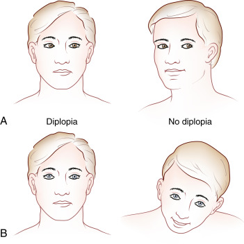
Diplopia is the perception of two images of a single object that may be displaced horizontally, vertically, or obliquely. Diplopia may be monocular or binocular. Monocular diplopia is double vision in one eye and persists when the unaffected eye is covered. It is an ophthalmologic problem that results from abnormal light transmission to the retina. Binocular diplopia results from ocular misalignment and resolves when either eye is covered. There are multiple possible causes that can be localized along the visual pathway, from the eye to the brainstem.
- Monocular diplopia: Persists when the unaffected eye is covered and, unless caused by a retinal defect, resolves when a pinhole is used.
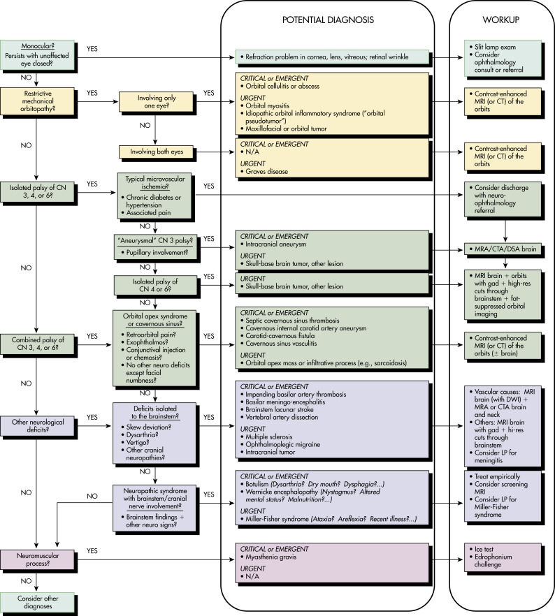
- Restrictive, mechanical orbitopathy: Restriction of eye movement away from affected muscle, typically gradual in onset with a sensation of mass effect/discomfort/pain in a single eye. It may be the result of rectus muscle entrapment secondary to orbital blowout fracture or may be accompanied by fever if infectious in etiology. When caused by thyroid disease, it may be unilateral or bilateral; be worse in the morning; and may have notable proptosis, periorbital swelling, edema, palpebral swelling, and conjunctival or scleral injection. Depending on the pathology, the patient may complain of pain.
- Cranial nerve (CN; III, IV, or VI) palsy: May occur as an isolated nerve palsy as a result of microvascular ischemia, or in combination in the case of cavernous sinus, central, or posterior orbital pathologies.
- CN III (oculomotor) palsy: Vertical and horizontal diplopia. Complete CN III palsy will have ptosis; large unreactive pupil; and paralysis of adduction, elevation, and depression so eye rests in position of abduction, depression, and intorsion.
- CN IV (trochlear) palsy: Vertical and/or torsional diplopia that worsens looking down and toward the nose; difficulty descending stairs, reading, or watching television in bed.
- CN VI (abducens) palsy: Horizontal diplopia that worsens on lateral gaze toward the affected side.
- Neuraxial process: D iplopia may occur in isolation or may be accompanied by multiple neurologic signs and symptoms depending on the location of the lesion and specific pathology.
- Neuromuscular disorder: Muscle atrophy/weakness, weakness on forced eyelid closure, normal reflexes, and no sensory deficits.
- The etiology of diplopia differs between monocular and binocular diplopia. Monocular diplopia (Box E1) is caused by primary ophthalmologic structural problems in the transmission of light to the retina and generally should be referred to an ophthalmologist. Binocular diplopia, when not caused by trauma, is most often a vascular lesion (small-vessel vasculopathy or aneurysmal disease) or occasionally due to endocrinopathy, neuromuscular, neoplastic, or autoimmune sources.
- Table E1 summarizes important causes of diplopia.
TABLE E1 Important Causes of Diplopia
| Diplopia-Causing Entity | Mechanism and Mortality | Distinguishing Features |
|---|---|---|
| Tier 1-Critical | ||
| Basilar artery thrombosis | Impending thrombosis of the basilar artery with brainstem ischemia; untreated mortality, 70%-90% | Vertigo, dysarthria, other cranial nerve involvement; risk factors for stroke |
| Botulism | Toxin inhibits release of acetylcholine (ACh) at cholinergic synapses and presynaptic myoneural junctions; untreated mortality, 60% | Dysarthria, dysphagia, autonomic dysreflexia, pupillary dysfunction |
| Basilar meningitis | Infection; untreated mortality, close to 100% if bacterial (15%-20% if treated) | Headache, meningismus, fever |
| Aneurysm | Enlarging aneurysm directly compresses cranial nerve; untreated rupture risk = 1%/yr (3.5%/yr for previously ruptured); mortality, 26%-67%/rupture | CN III palsy with pupillary involvement |
| Tier 2-Emergent | ||
| Vertebral dissection | Dissection causes vertebrobasilar ischemia; acute untreated mortality, 28% (2%-5% if neurologically asymptomatic) | Neck pain, vertigo; risk factors for vertebral dissection |
| Myasthenia gravis | Autoantibodies develop against ACh nicotinic postsynaptic receptors; untreated crisis mortality, 42% (5% if treated) | Fluctuating muscle weakness, ptosis, and diplopia worsen with activity and improve with rest |
| Wernicke encephalopathy | Thiamine-dependent metabolic failure and tissue injury; untreated mortality, 20% | Nystagmus, ataxia, altered mental status, and ophthalmoplegia; alcoholism and nutritional deficiency |
| Orbital apex syndrome, cavernous sinus process | Inflammation or infection in the orbital apex or cavernous sinus directly affects oculomotor cranial nerves; acute mortality low unless infectious and complicated by meningitis | A combination of palsies of CN III, IV, or VI, with retro-orbital pain, conjunctival injection, possible periorbital, facial numbness |
| Tier 3-Urgent | ||
| Brainstem tumor | Tumor involvement at the supranuclear level; acute mortality low (long-term mortality variable) | Skew deviation-vertical diplopia, internuclear ophthalmoplegia |
| Miller-Fisher syndrome | Autoantibodies develop to a cranial nerve ganglioside, GQ1b; acute mortality low (if fully differentiated from GBS; mortality, 2%-12% if GBS) | Ophthalmoplegia, ataxia, areflexia |
| Multiple sclerosis | Demyelinating lesions; acute mortality low | Internuclear ophthalmoplegia |
| Thyroid myopathy (Graves’ disease) | Autoimmune myopathy; acute mortality low in regard to ocular complaints | Proptosis, restriction of elevation and abduction of the eye, signs of Graves’ disease |
| Ophthalmoplegic migraine | Inflammatory cranial neuropathy; low mortality, self-limited disease | Ipsilateral headache, CN (usually III) palsy |
| Ischemic neuropathy | Microvascular ischemia; mortality low, self-limited disease | Isolated CN palsy (pupil-sparing if CN III) |
| Orbital myositis, pseudotumor | Autoimmune or idiopathic myositis; acute mortality low in regard to ocular complaints | Eye pain, restriction of movement, periorbital edema; exophthalmos and chemosis when more severe |
| Orbital apex mass | Tumor, infiltration, or mass effect in orbital apex or cavernous sinus directly compresses oculomotor cranial nerves; acute mortality low | A combination of palsies of CN III, IV, or VI, and possible periorbital, facial numbness, with retro-orbital pain, proptosis, signs of venous congestion |
CN, Cranial nerve; GBS, Guillain-Barré syndrome.
From Walls RM et al: Rosen’s emergency medicine, ed 9, Philadelphia, 2018, Elsevier.
BOX E1 Causes of Monocular Diplopia
From Jankovic J et al: Bradley and Daroff’s neurology in clinical practice, ed 8, Philadelphia, 2022, Elsevier.