| Author: Victoria Wilson
|
| Author: Victoria Wilson
|
A Look at Acid-Base Imbalances
The body continually works to maintain a balance (homeostasis) between acids and bases. Without that balance, cells can't function properly. As cells use nutrients to produce the energy they need to function, two by-products are formed—carbon dioxide and hydrogen. Acid-base balance depends on the regulation of free hydrogen ions. The concentration of hydrogen ions in body fluids determines the extent of acidity or alkalinity, both of which are measured in pH. Remember, pH levels are inversely proportionate to hydrogen ion concentration, which means that when hydrogen concentration increases, pH decreases (acidosis). Conversely, when hydrogen concentration decreases, pH increases (alkalosis). (See chapter 3, Balancing acids and bases.)
Gas gives good answers
Blood gas measurements remain the principal diagnostic tool for evaluating acid-base states. An arterial blood gas (ABG) analysis includes these tests: pH, which measures the hydrogen ion concentration and is an indication of the blood's acidity or alkalinity; partial pressure of arterial carbon dioxide (PaCO2), which reflects the adequacy of ventilation by the lungs; and bicarbonate level, which indicates the activity of the kidneys in retaining or excreting bicarbonate. (See The ABCs of ABGs.)
Normal fix-me-ups
Most of the time, the body's compensatory mechanisms restore acid-base balance—or at least prevent the life-threatening conse quences of an imbalance. Those compensatory mechanisms include chemical buffers as well as certain respiratory and kidney reactions.
For example, the body compensates for a primary respiratory disturbance such as respiratory acidosis by inducing metabolic alkalosis. Unfortunately, not all attempts to compensate are equal. The respiratory system is efficient and can compensate for metabolic disturbances quickly, whereas the metabolic system, working through the kidneys, can take hours or days to compensate for an imbalance. Therefore, intervention is necessary to avoid further patient deterioration. This chapter takes a closer look at each of the four major acid-base imbalances.
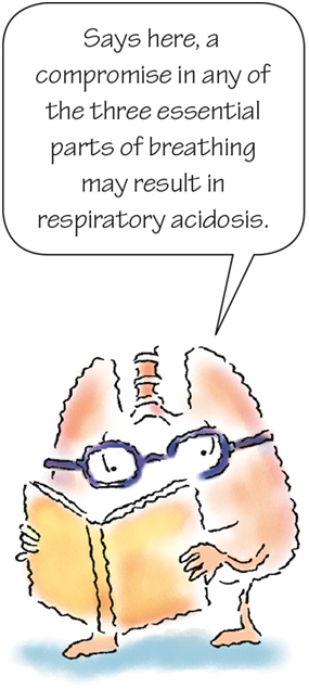
A compromise in any of the three essential parts of breathing— ventilation, perfusion, or diffusion—may result in respiratory acidosis. This acid-base disturbance is characterized by alveolar hypoventilation, meaning the pulmonary system is unable to rid the body of enough carbon dioxide to maintain a healthy pH balance. This reaction occurs as a result of decreased respiration or inadequate gas exchange.
The lack of efficient carbon dioxide release leads to hypercapnia in which PaCO2 is greater than 45 mm Hg. The condition can be acute, resulting from a sudden failure in ventilation, or chronic, resulting from chronic pulmonary disease.
In acute respiratory acidosis, pH drops below normal (lower than 7.35). In chronic respiratory acidosis, commonly due to chronic obstructive pulmonary disease (COPD), pH stays within normal limits (7.35 to 7.45) because the kidneys have had time to compensate for the imbalance. (More on that complex phenomenon later.)
How it happens
When a patient hypoventilates, carbon dioxide builds up in the bloodstream, and pH drops below normal—respiratory acidosis. The kidneys try to compensate for a drop in pH by conserving bicarbonate (base) ions or generating them in the kidneys, which in turn raises the pH. (See What happens in respiratory acidosis.)
Respiratory acidosis can result from neuromuscular problems, depression of the respiratory center in the brain, lung disease, obesity, postoperative pain, or airway obstruction.
That breathless feeling
In certain neuromuscular diseases—such as Guillain-Barré syndrome, myasthenia gravis, and poliomyelitis—the respiratory muscles fail to respond appropriately to the respiratory drive, resulting in respiratory acidosis. Diaphragmatic paralysis, which commonly occurs with spinal cord injury, works the same way to cause respiratory acidosis.
Hypoventilation from CNS trauma or brain lesions—such as tumors, vascular disorders, or infections—may impair the patient's ventilatory drive. Obesity (as in pickwickian syndrome) or primary hypoventilation (as in Ondine curse) may contribute to this imbalance as well. Also, certain medications—including anesthetics, hypnotics, opioids, and sedatives—can depress the respiratory center of the brain, leading to hypercapnia. (See Medications associated with respiratory acidosis.)
Scanty surface
Lung diseases that decrease the amount of pulmonary surface area available for gas exchange can prompt respiratory acidosis. Less surface area reduces the amount of gas exchange that can occur, thus impeding carbon dioxide exchange. Examples of pulmonary problems that can decrease surface area include respiratory infections, COPD, acute asthma attacks, chronic bronchitis, late stages of adult respiratory distress syndrome, pulmonary edema, conditions in which there is increased dead space in the lungs (hypoventilation), and physiologic or anatomic shunts.
Chest wall trauma (leading to pneumothorax or flail chest) can also cause respiratory acidosis. The ventilatory drive remains intact, but the chest wall mechanics of the collapsed lung don't allow for enough alveolar ventilation to meet the body's needs. Chest wall mechanics can be impeded as a result of the rib cage distortion caused by fibrothorax or kyphoscoliosis.
Danger! Obstruction ahead
Respiratory acidosis can also be caused by airway obstruction, which leads to carbon dioxide retention in the lungs. Airway obstruction can occur as a result of retained secretions, tumors, anaphylaxis, laryngeal spasm, or lung diseases that interfere with alveolar ventilation. Keep in mind that children are particularly prone to airway obstruction, as are older adults and patients who are debilitated, who may not be able to effectively clear secretions. Other factors also increase an infant's risk of developing acidosis. (See Infants and acidosis.)
Risky business
Treatments can also induce respiratory acidosis. For instance, mechanical ventilation that under ventilates a patient can cause carbon dioxide retention. A postoperative patient is at risk for respiratory acidosis if fear of pain prevents the patient from participating in pulmonary hygiene measures, such as using the incentive spirometer and coughing and deep breathing. Also, analgesics or sedatives can depress the medulla (which controls respirations), leading to inadequate ventilation and subsequent respiratory acidosis.
What to look for
Signs and symptoms of respiratory acidosis depend on the cause of the condition. The patient may report a headache because carbon dioxide dilates cerebral blood vessels. (See Signs and symptoms of respiratory acidosis.)
CNS depression may result in an altered LOC, ranging from restlessness, confusion, and apprehension to lethargy and coma. If acidosis remains untreated, a fine flapping tremor and depressed reflexes may develop. The patient may also report nausea and vomiting, and the skin may be warm and flushed.
A breakdown in breathing
Most patients with respiratory acidosis have rapid, shallow respirations; they may be dyspneic and diaphoretic. Auscultation reveals diminished or absent breath sounds over the affected area. However, if acidosis stems from CNS trauma or lesions or drug overdose, the respiratory rate is significantly decreased.
In a patient with acidosis, hyperkalemia, and hypoxemia, you may note tachycardia and ventricular arrhythmias. Cyanosis is a late sign of the condition. Resulting myocardial depression may lead to shock and, ultimately, cardiac arrest.
What tests show
Several test results help confirm a diagnosis of respiratory acidosis and guide treatment:
How it's treated
Treatment of respiratory acidosis focuses on improving ventilation and lowering PaCO2. If respiratory acidosis stems from nonpulmonary conditions, such as neuromuscular disorders or a drug overdose, treatment aims to correct or improve the underlying cause.
Treatment for respiratory acidosis with a pulmonary cause includes:
How you intervene
If your patient develops respiratory acidosis, maintain a patent airway. Help remove any foreign bodies from the patient's airway and establish an artificial airway. Provide adequate humidification to keep the patient's secretions moist. Also, follow these measures:

Oxygen: Too much of a good thing
As you reevaluate your patient's condition, consider these questions:
The opposite of respiratory acidosis, respiratory alkalosis results from alveolar hyperventilation and hypocapnia. In respiratory alkalosis, increased elimination of carbon dioxide occurs; therefore, pH is greater than 7.45 and PaCO2 is less than 35 mm Hg. Acute respiratory alkalosis results from a sudden increase in ventilation. Chronic respiratory alkalosis may be challenging to identify because of renal compensation.
How it happens
Any clinical condition that increases respiratory rate or depth can cause the lungs to eliminate, or “blow off,” carbon dioxide. Because carbon dioxide is an acid, eliminating it causes a decrease in PaCO2 along with an increase in pH—alkalosis.
Hyperventilation (gasp!)
The most common cause of acute respiratory alkalosis is hyperventilation stemming from an anxiety or panic attack. It may also occur during cardiopulmonary resuscitation when rescuers hyperventilate the patient at 30 to 40 breaths per minute. Pain can have the same effect. Hyperventilation is also an early sign of salicylate intoxication and can occur with the use of nicotine, xanthines such as aminophylline, and other medications. (See Medications associated with respiratory alkalosis.)
Hypermetabolic states—such as fever, liver failure, and sepsis (especially gram-negative sepsis)—can lead to respiratory alkalosis. Conditions that affect the respiratory control center in the medulla are also a danger. For example, the higher progesterone levels during pregnancy may stimulate this center, whereas stroke or trauma may injure it, both resulting in respiratory alkalosis.
Hypoxia (pant!)
Acute hypoxia, secondary to high altitude, pulmonary disease, severe anemia, pulmonary embolus, or hypotension, can cause respiratory alkalosis. Such conditions may overstimulate the respiratory center and cause the patient to breathe faster and deeper. Overventilation during mechanical ventilation causes the lungs to blow off more carbon dioxide, resulting in respiratory alkalosis.
What to look for
An increase in the rate and depth of respiration is a primary sign of respiratory alkalosis. It's also common for the patient to have tachycardia. The patient may appear anxious and restless as well as complain of light-headedness, muscle weakness, fear, or difficulty breathing. (See What happens in respiratory alkalosis.)
In extremis
In extreme alkalosis, confusion or syncope may occur. Because of the lack of carbon dioxide in the blood and its effect on cerebral blood flow and the respiratory center, you may see alternating periods of apnea and hyperventilation. The patient may report tingling in the fingers and toes.
ECG exposé
You may see ECG changes, including a prolonged PR interval, a flattened T wave, a prominent U wave, and a depressed ST segment. (For more information, see chapter 6, When potassium tips the balance.)

Signs of trouble
Signs and symptoms worsen as calcium levels drop because of vasoconstriction of peripheral and cerebral vessels resulting from hypoxia. You may see hyperreflexia, carpopedal spasm, tetany, arrhythmias, a progressive decrease in the patient's LOC, seizures, or coma. (See Signs and symptoms of respiratory alkalosis.)
What tests show
Several diagnostic test results may help detect respiratory alkalosis and guide treatment:
How it's treated
Treatment focuses on correcting the underlying disorder, which may require removing the causative agent (such as salicylate or other medications), taking steps to reduce fever and eliminate the source of sepsis. If acute hypoxemia is the cause, the patient will need oxygen therapy. If anxiety is the cause, the patient may receive a sedative or an anxiolytic. If the patient is having pain, the patient may receive an analgesic.
It's in the bag
To counteract hyperventilation, the patient can breathe into a paper bag or cupped hands. As a result, the patient breathes exhaled carbon dioxide, thereby raising the carbon dioxide level. If a patient's respiratory alkalosis is medically induced, mechanical ventilator settings may be adjusted by decreasing the tidal volume or the number of breaths per minute.
How you intervene
Monitor patients at risk of developing respiratory alkalosis. If your patient develops the condition, take these actions:
Metabolic acidosis is caused by an increase in hydrogen ion production and is characterized by a pH below 7.35 and a bicarbonate level below 22 mEq/L. This disorder depresses the CNS. Left untreated, it may lead to ventricular arrhythmias, coma, and cardiac arrest.
How it happens
The underlying mechanisms in metabolic acidosis are a loss of bicarbonate from extracellular fluid, the accumulation of metabolic acids, or a combination of the two. If the patient's anion gap (measurement of the difference between the amount of sodium and the amount of bicarbonate in the blood) is greater than 14 mEq/L, then the acidosis is a result of an accumulation of metabolic acids (unmeasured anions).
If metabolic acidosis is associated with a normal anion gap (8 to 14 mEq/L), loss of bicarbonate may be the cause. (See What happens in metabolic acidosis.)
Acids ante up; bases bottom out
Metabolic acidosis is characterized by a gain in acids or a loss of bases from the plasma and may be related to an overproduction of ketone bodies. When the body has used up its glucose supplies, it draws on its fat stores for energy, converting fatty acids to ketone bodies. Conditions that cause an overproduction of ketone bodies include diabetes mellitus, chronic alcoholism, severe malnutrition or starvation, inadequate dietary intake of carbohydrates, hyperthyroidism, and acute infection with accompanying fever.
Lactic acidosis can cause or worsen metabolic acidosis and can occur secondary to shock, heart failure, pulmonary disease, hepatic disorders, seizures, or strenuous exercise.
Kidney culprit
Metabolic acidosis can also stem from a decreased ability of the kidneys to excrete acids, as occurs in renal insufficiency or kidney failure with acute tubular necrosis.
Gut reactions
Metabolic acidosis also occurs with excessive gastrointestinal (GI) losses from diarrhea, intestinal malabsorption, a draining fistula of the pancreas or liver, or a urinary diversion to the ileum. Other causes include hyperaldosteronism and use of a potassium-sparing diuretic such as acetazolamide, which inhibits the secretion of acid.
Poison pills
Patients with poisoning or a toxic reaction to a medication are at particular risk for metabolic acidosis. This can occur following inhalation of toluene or ingestion of a salicylate (such as an aspirin or aspirin-containing medication), methanol, ethylene glycol, paraldehyde, hydrochloric acid, or ammonium chloride.

What to look for
Metabolic acidosis typically produces respiratory, neurologic, and cardiac signs and symptoms. As acid builds up in the bloodstream, the lungs compensate by blowing off carbon dioxide.
Hyperventilation (especially increased depth of respiration) is the first clue to metabolic acidosis. Called Kussmaul respirations, the breathing is rapid and deep. A patient with diabetes who experiences Kussmaul respirations may have a fruity breath odor. The odor stems from catabolism of fats and excretion of acetone through the lungs.
So depressing
As pH drops, the CNS is further depressed, as is the myocardial function. Cardiac output and blood pressure drop, and arrhythmias may occur if the patient also has hyperkalemia.
Initially, the skin is warm and dry as a result of peripheral vasodilation, but, as shock develops, the skin becomes cold and clammy. As the cerebral vessels dilate, the patient may complain of weakness and a dull headache.
The patient's LOC may deteriorate from confusion to stupor and coma. A neuromuscular examination may show diminished muscle tone and deep tendon reflexes. Metabolic acidosis also affects the GI system, causing anorexia, nausea, and vomiting. (See Signs and symptoms of metabolic acidosis.)
What tests show
Several test results may help diagnose and treat metabolic acidosis:
How it's treated
Treatment aims to correct the acidosis as quickly as possible by addressing both the symptoms and the underlying cause. Respiratory compensation is usually the first line of therapy, including mechanical ventilation if needed.
Put potassium in its place
For patients with diabetes, expect to administer rapid-acting insulin to reverse DKA and drive potassium back into the cell. For any patient with metabolic acidosis, monitor serum potassium levels. Even though the patient initially has high serum potassium levels, the levels drop as acidosis is corrected, and the patient may end up with hypokalemia. Any other electrolyte imbalances should be evaluated and corrected.
Bump up the bicarbonate
Expect to administer IV sodium bicarbonate to neutralize blood acidity in patients with bicarbonate loss and a pH lower than 7.1. Fluids are replaced parenterally, as required. Dialysis may be initiated in patients with kidney failure or a toxic reaction to a medication. Such patients may receive an antibiotic to treat sources of infection or an antidiarrheal to treat diarrhea-induced bicarbonate loss.
Always on the alert
Watch for signs of worsening CNS status or deteriorating laboratory and ABG test results. The patient may need ventilatory support, so prepare for intubation. A patient with kidney failure may require dialysis, especially when this condition is complicated by diabetes. Maintain a patent IV line to administer emergency medications. Flush the line with normal saline solution before and after administering sodium bicarbonate because the bicarbonate may inactivate or cause precipitation of many medications. (See Acidosis and dopamine.)
How you intervene
If your patient is at risk for metabolic acidosis, careful monitoring can help prevent it from developing.
If your patient already has metabolic acidosis, nursing care includes immediate emergency interventions and long-term treatment of the condition and its underlying causes. Observe the following guidelines:
Q & A time
Physical examination and further diagnostic tests may provide more information about your patient's metabolic acidosis. As you reevaluate the patient's condition, consider these questions:
Metabolic alkalosis is caused by a decrease in hydrogen ion production, characterized by a blood pH above 7.45, and accompanied by a bicarbonate level above 26 mEq/L. In acute metabolic alkalosis, bicarbonate may be as high as 50 mEq/L. With early diagnosis and prompt treatment, the prognosis for effective treatment is favorable. Untreated, metabolic alkalosis can result in coma, arrhythmias, and death.
How it happens
In metabolic alkalosis, the underlying mechanisms include a loss of hydrogen ions (acid), a gain in bicarbonate, or both. A PaCO2 level greater than 45 mm Hg (possibly as high as 60 mm Hg) indicates that the lungs are compensating for alkalosis. Renal compensation is more effective but slower. Metabolic alkalosis is commonly associated with hypokalemia, particularly from the use of thiazides, furosemide, ethacrynic acid, and other diuretics that deplete potassium stores. In hypokalemia, the kidneys conserve potassium. At the same time, the kidneys also increase the excretion of hydrogen ions, which prompts alkalosis from the loss of acid. Metabolic alkalosis may also occur with hypochloremia and hypocalcemia. (See What happens in metabolic alkalosis.)
GI grief
Metabolic alkalosis can result from many causes, the most common of which is an excessive acid loss from the GI tract. Vomiting causes loss of hydrochloric acid from the stomach. Children who have pyloric stenosis can develop this disorder. Alkalosis also results from prolonged nasogastric (NG) suctioning, presenting a risk for surgical patients and patients with GI disorders.
Diuretic danger
Diuretic therapy presents another risk of metabolic alkalosis. Thiazide and loop diuretics can lead to a loss of hydrogen, potassium, and chloride ions from the kidneys. Hypokalemia causes the kidneys to excrete hydrogen ions as they try to conserve potassium. Potassium moves out of the cells as hydrogen moves in, resulting in alkalosis.
With the fluid loss from diuresis, the kidneys attempt to conserve sodium and water. For sodium to be reabsorbed, hydrogen ions must be excreted. In a process known as contraction alkalosis, bicarbonate is reabsorbed and metabolic alkalosis results.
More metabolic mishaps
Cushing disease can lead to metabolic alkalosis by causing retention of sodium and chloride and urinary loss of potassium and hydrogen. Rebound alkalosis following correction of organic acidosis, such as after cardiac arrest and administration of sodium bicarbonate, can also cause metabolic alkalosis. Posthypercapnic alkalosis occurs when chronic carbon dioxide retention is corrected by mechanical ventilation, and the kidneys haven't yet corrected the chronically high bicarbonate levels.
Metabolic alkalosis can also result from kidney diseases, such as renal artery stenosis, or multiple transfusions. Certain medications, such as corticosteroids and antacids that contain sodium bicarbonate, can also lead to metabolic alkalosis. (See Medications associated with metabolic alkalosis.) Continuous NG tube suction can also cause metabolic alkalosis due to the removal of acid and electrolytes.
What to look for
Initially, your patient may have slow, shallow respirations as hypoventilation, a compensatory mechanism, occurs. However, this mechanism is limited because hypoxemia soon develops, which stimulates ventilation. The signs and symptoms of metabolic alkalosis are commonly associated with an underlying condition. Characteristic hypokalemic or hypocalcemic ECG changes may occur as well as signs of hypotension.
A neurologic nightmare
Metabolic alkalosis results in neuromuscular excitability, which causes muscle twitching, weakness, and tetany. The patient develops hyperactive reflexes and may also experience numbness and tingling of the fingers, toes, and mouth area. Neurologic symptoms include apathy and confusion. Seizures, stupor, and coma may result if severe.
Keep track of these tracts
If hypokalemia affects the GI tract, the patient is likely to experience anorexia, nausea, and vomiting. If it affects the genitourinary (GU) tract—that is, if the kidneys are affected—polyuria may result. If left untreated, metabolic alkalosis can result in arrhythmias and death. (See Signs and symptoms of metabolic alkalosis.)
What tests show
These tests may be helpful in the diagnosis and treatment of metabolic alkalosis:
How it's treated
Treatment aims to correct the acid-base imbalance by providing the patient's body enough time to rid itself of excess bicarbonate and increase its hydrogen concentration. Treatment may include:
How you intervene
If your patient is at risk for metabolic alkalosis, careful monitoring can help prevent its development.
If your patient already has metabolic alkalosis, follow these guidelines:
 If you answered all five questions correctly, wow! Test the pH of the nearest pool and jump in for a refreshing swim!
 If you answered four correctly, excellent! You're just about ready to do your first solo balancing act!
 If you answered fewer than four correctly, take heart. You're still a boffo buffer in our book. (Buffer, get it? For chemical buffers? Oh well, can't win 'em all.)
References
Alfano, G., Fontana, F., Mori, G., Giaroni, F., Ferrari, A., Giovanella, S., Ligabue, G., Ascione, E., Cazzato, S., Ballestri, M., Di Gaetano, M., Meschiari, M., Menozzi, M., Milic, J., Andrea, B., Franceschini, E., Cuomo, G., Magistroni, R., Mussini, C., … Guaraldi, G. (2021, June 11). Acid base disorders in patients with COVID-19. International Urology and Nephrology. https://link.springer.com/article/10.1007/s11255-021-02855-1
Bronfenbrener, R. (2020, September 24). Acid-base interpretation: Reference range, interpretation, collection and panels. https://emedicine.medscape.com/article/2058760-overview
Chawla, R., Dixit, S. B., Zirpe, K. G., Chaudhry, D., Khilnani, G. C., Mehta, Y., Khatib, K. I., Jagiasi, B. G., Chanchalani, G., Mishra, R. C., Samavedam, S., Govil, D., Gupta, S., Prayag, S., Ramasubban, S., Dobariya, J., Marwah, V., Sehgal, I., Jog, S. A., & Kulkarni, A. P. (2020, January 24). ISCCM guidelines for the use of non-invasive ventilation in acute respiratory failure in adult ICUs. Indian Journal of Critical Care Medicine. https://www.ncbi.nlm.nih.gov/pmc/articles/PMC7085817/
Hamm, L. L., Nakhoul, N., & Hering-Smith, K. S. (2015). Acid-base homeostasis. Clinical Journal of the American Society of Nephrology: CJASN, 10(12), 2232–2242. https://www.ncbi.nlm.nih.gov/pmc/articles/PMC4670772/
Hopkins, E., Sanvictores, T., & Sharma, S. (n.d.). Physiology, acid base balance – Statpearls – NCBI bookshelf. National Library of Medicine. https://www.ncbi.nlm.nih.gov/books/NBK507807/
Lippincott's visual nursing: A guide to disease skills and treatment (2nd ed.). (2012). Lippincott Williams & Wilkins.
Magder, S., & Emami, A. (2015). Practical approach to physical-chemical acid-base management: Stewart at the bedside. Annals of the American Thoracic Society, 12(1), 111–117. https://www.atsjournals.org/doi/full/10.1513/AnnalsATS.201409-426OI?url_ver=Z39.88-2003&rfr_id=ori%3Arid%3Acrossref.org&rfr_dat=cr_pub%3Dpubmed
Snyder, L. M., Rao, L. V., & Wallach, J. B. (2021). Wallach's interpretation of diagnostic tests (11th ed.). Lippincott Williams & Wilkins.
Sultana, R., Ahsan, A. S. M. A., Fatema, K., Ahmed, F., Saha, D. K., Saha, M., Nazneen, S., Mahbub, N., & Ashraf, E. (2020, December 27). Pattern of electrolytes in a cohort of critically ill COVID-19 patients. BIRDEM Medical Journal. https://www.banglajol.info/index.php/BIRDEM/article/view/50980