section name header

Basic Information ⬇

Author: Philip A. Chan, MD, MS

Definition

Acquired immunodeficiency syndrome (AIDS) is a disorder caused by infection with HIV and marked by progressive deterioration of the cellular immune system, leading to secondary (opportunistic) infections and/or malignancies. AIDS-defining conditions are summarized in Box 1.

BOX 1 AIDS-Defining Conditions

  • •Bacterial infections, multiple or recurrent
  • •Candidiasis of bronchi, trachea, or lungs
  • •Candidiasis of esophagus
  • •Cervical cancer, invasive
  • •Coccidioidomycosis, disseminated or extrapulmonary
  • •Cryptococcosis, extrapulmonary
  • •Cryptosporidiosis, chronic intestinal (>1 mo duration)
  • •Cytomegalovirus disease (other than liver, spleen, or nodes), onset at age >1 mo
  • •Cytomegalovirus retinitis (with loss of vision)
  • •Encephalopathy, HIV related
  • •Herpes simplex: chronic ulcers (>1 mo duration) or bronchitis, pneumonitis, or esophagitis (onset at age >1 mo)
  • •Histoplasmosis, disseminated or extrapulmonary
  • •Isosporiasis, chronic intestinal (>1 mo duration)
  • •Lymphoma, Burkitt (or equivalent term)
  • •Kaposi sarcoma
  • •Lymphoma, immunoblastic (or equivalent term)
  • •Lymphoma, primary, of brain
  • •Mycobacterium avium complex or Mycobacterium kansasii, disseminated or extrapulmonary
  • •Mycobacterium tuberculosis of any site, pulmonary, disseminated, or extrapulmonary
  • •Mycobacterium, other species or unidentified species, disseminated or extrapulmonary
  • •Pneumocystis jirovecipneumonia
  • •Pneumonia, recurrent
  • •Progressive multifocal leukoencephalopathy
  • •Salmonella septicemia, recurrent
  • •Toxoplasmosis of brain, onset at age >1 mo
  • •Wasting syndrome attributed to HIV

From Walls RM et al: Rosen’s emergency medicine, concepts and clinical practice, ed 10, Philadelphia, 2023, Elsevier.

Synonym

  • AIDS
ICD-10CM CODE
B20Human immunodeficiency virus [HIV] disease
Epidemiology & Demographics
Incidence (In U.S.):

  • •A significant number of persons are diagnosed with AIDS in the U.S. each year.
  • •There is a disproportionate number of new HIV/AIDS cases among Black/African Americans and Latino/Hispanic Americans compared with White Americans.
  • •The majority of all new HIV/AIDS diagnoses are among gay, bisexual, or other men who have sex with men (MSM).
Prevalence (In U.S.):

Approximately 1.2 million people in the U.S. have HIV. A subset of these have been diagnosed with AIDS.

Predominant Sex:

Men constitute the majority of HIV/AIDS diagnoses in the U.S. with a disproportionate number among MSM.

Predominant Age:

The predominant age group diagnosed with HIV/AIDS is 25 to 59 yr of age.

Peak Incidence:

Ages 25 to 59 yr

Genetics:

  • •Familial disposition: Although there is no proven genetic predisposition, individuals with deletions in the CCR5 gene are less susceptible to HIV infection with macrophage tropic virus (the predominant virus in sexual transmission) and may progress to AIDS more slowly.
  • •Congenital infection:
    1. 1.HIV is transmittable from an infected mother to the fetus in utero in as many as 30% of pregnancies if untreated.
    2. 2.No specific congenital malformations associated with infection; low birth weight and spontaneous abortion are possible.
Physical Findings & Clinical Presentation

  • •Nonspecific findings: Fever, weight loss, anorexia
  • •Specific syndromes:
    1. 1.Seen in association with specific opportunistic infections and malignancies. These include:
      1. a.Opportunistic infections:
        • (1)Disseminated strongyloidiasis
        • (2)Disseminated toxoplasmosis, cryptococcosis, histoplasmosis, cytomegalovirus (CMV), herpes simplex virus (HSV), or mycobacterial disease (most common is Mycobacterium avium complex)
        • (3)Candida esophagitis or bronchopulmonary disease
        • (4)Chronic cryptosporidiosis diarrhea
        • (5)Pneumocystis jiroveci pneumonia (PJP)
        • (6)Extensive pulmonary and extra-pulmonary tuberculosis (TB)
        • (7)Recurrent pneumonia or other bacterial infections
        • (8)Progressive multifocal leukoencephalopathy (PML)
        • (9)Cutaneous lesions (Box 2)
      2. b.AIDS-related neoplasms:
        • (1)Kaposi sarcoma
        • (2)Primary brain lymphoma
        • (3)Invasive cervical carcinoma
        • (4)High-grade B-cell non-Hodgkin lymphoma, Burkitt lymphoma, undifferentiated non-Hodgkin lymphoma, or immunoblastic lymphoma
    2. 2.Most common:
      1. a.Respiratory infections (Pneumocystis jiroveci pneumonia, TB, bacterial pneumonia, fungal infection [Table 1])
      2. b.CNS infections (toxoplasmosis, TB [Table 2])
      3. c.GI (cryptosporidiosis, isosporiasis, CMV); Sections II and III describe organisms associated with diarrhea in patients with AIDS
      4. d.Eye infections (CMV, toxoplasmosis)
      5. e.Kaposi sarcoma (cutaneous or visceral) or lymphoma (nodal or extranodal)
  • •Possibly asymptomatic
  • •Diagnosis of AIDS if the CD4 cell count is <200 or <14% of total lymphocyte in the presence of proven HIV infection, even in the absence of other infections
  • •The various manifestations of HIV infection are described in Section II

BOX 2 Cutaneous Findings Highly Suggestive of HIV Disease

  • •Any WHO criteria for stage 4 HIV disease
  • •Facial molluscum in an adult
  • •Proximal subungual onychomycosis
  • •Herpes zoster scarring
  • •Oral hairy leukoplakia
  • •Bacillary angiomatosis
  • •Widespread dermatophytosis
  • •Severe seborrheic dermatitis

From Walls RM et al: Rosen’s emergency medicine, concepts and clinical practice, ed 10, Philadelphia, 2023, Elsevier.

TABLE 1 Differential Diagnosis of Respiratory Infections in HIV-Infected Patients by CD4+ Count

CD4+ Count and StageDifferential Diagnosis
Present at any stageAcute bronchitis
Bacterial pneumonia
Tuberculosis
>500 cells/μLBacterial pneumoniaa
Early HIV infectionPCPa
HHV-8-related Kaposi sarcoma
200-500 cells/μLBacterial pneumoniaa
PCPa
<200 cells/μLBacterial pneumoniaa (consider bacteremia)
AIDSPCPa
Histoplasma capsulatum or Coccidioides immitis pneumonia
Cryptococcus neoformans pneumonia
Extrapulmonary or disseminated tuberculosisa
≤50 cells/μLBacterial pneumoniaa
Advanced HIV infectionPCPa
Toxoplasma gondii pneumonia
Pulmonary Kaposi sarcoma
Histoplasma capsulatum or Coccidioides immitis pneumonia
Mycobacterium avium complex pneumonia

HHV-8, Human herpesvirus 8; PCP, Pneumocystis jiroveci pneumonia.

a Occurs more frequently as immune function declines.

From Walls RM et al: Rosen’s emergency medicine, concepts and clinical practice, ed 10, Philadelphia, 2023, Elsevier.

TABLE 2 Differential of Focal Central Nervous System Lesions in Patients With HIV Infection

Common Clinical PresentationImaging and Diagnostic Testing
Toxoplasma encephalitis
  • Fever
  • Headache
  • Altered mental status
  • Focal neurologic findings
  • Seizure
  • Evolves during days to weeks
  • Ring enhancing (≈90% of the time) CNS lesions
  • Frequent edema and mass effect
  • Toxoplasma antibodies (reflects past exposure)
  • CD4+ often <100 cells/μL
  • PCR detection of Toxoplasma gondii
Primary CNS lymphoma (PCNSL)
  • Confusion
  • Lethargy
  • Memory loss
  • Hemiparesis
  • Aphasia
  • Seizure
  • Fever
  • Night sweats
  • Weight loss
  • Evolves during months
  • CNS lesion or lesions (may have mass effect)
  • Solitary lesions are often large (>4 cm)
  • Some ring enhancement may occur but less regular
  • PCR assay for Epstein-Barr virus (associated with PCNSL)
Progressive multifocal leukoencephalopathy (PML)
  • Progressive focal neurologic deficits (during months)
  • Hemiparesis
  • Visual field defects
  • Ataxia
  • Aphasia
  • Cognitive impairment
  • Multifocal areas of demyelination primarily involving white matter
  • Less frequent mass effect or ring-enhancing
  • PCR assay for DNA of JC virus (causes PML)
HIV encephalopathy
  • Memory and psychomotor speed impairment
  • Depressive symptoms
  • Movement disorders
  • Multiple hyperintense signals in T2-weighted images
  • Often symmetric; not well demarcated
Cytomegalovirus encephalitis
  • Delirium
  • Confusion
  • Focal neurologic abnormalities
  • Magnetic resonance imaging shows multifocal scattered micronodules and ventriculoencephalitis.
  • CD41+ <50 cells/μL
Brain abscess
  • Focal neurologic deficit
  • Headache
  • Bacteremia or craniofacial infection
  • Often concomitant evidence of disseminated infection
Tuberculoma
  • Focal neurologic deficit
  • Headache
  • Tuberculosis infection
  • Single or multiple mass lesions
  • Can be manifested as focal lesion or meningeal infection

CNS, Central nervous system; PCR, polymerase chain reaction.

From Walls RM et al: Rosen’s emergency medicine, concepts and clinical practice, ed 10, Philadelphia, 2023, Elsevier.

Etiology

  • •Caused by infection with HIV-1 or HIV-2 (less common).
  • •HIV is transmitted by sexual contact, needle-sharing (during injection drug use), transfusion of contaminated blood or blood products, and from infected mother to fetus or neonate.

Diagnosis ⬆ ⬇

Differential Diagnosis

  • •Other wasting illnesses mimicking the nonspecific features of AIDS:
    1. 1.TB
    2. 2.Malignancy
    3. 3.Disseminated fungal infection
    4. 4.Malabsorption syndromes
    5. 5.Depression
  • •Other disorders associated with dementia or demyelination producing encephalopathy, myelopathy, or neuropathy
Workup

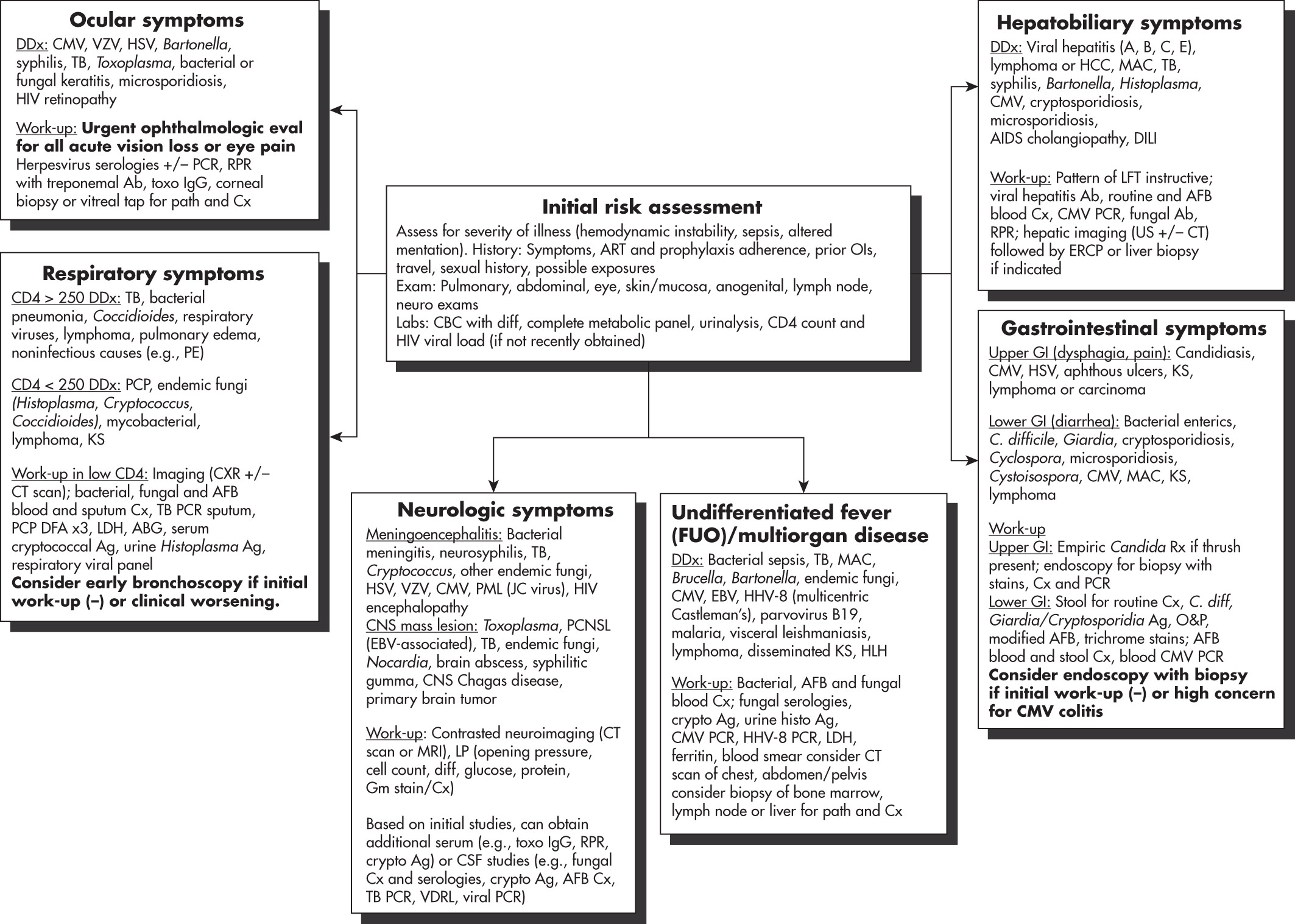
Prompt evaluation of respiratory, CNS, and GI complaints. Fig. 1 illustrates a syndromic approach to suspected opportunistic infection in HIV.

Figure 1 Syndromic approach to suspected opportunistic infection in HIV.

!!flowchart!!

ABG, Arterial blood gas; AFB, acid-fast bacilli; ART, antiretroviral therapy; CBC, complete blood count; CMV, cytomegalovirus; DFA, direct fluorescent antibody; DILI, drug-induced liver injury; EBV, Epstein-Barr virus; ERCP, endoscopic retrograde cholangiopancreatography; FUO, fever of unknown origin; HCC, hepatocellular carcinoma; HHV-8, human herpes virus 8; HLH, hemophagocytic lymphohistiocytosis; HSV, herpes simplex virus; KS, Kaposi sarcoma; LDH, lactate dehydrogenase; LP, lumbar puncture; MAC, Mycobacterium avium complex; O&P, ova and parasite; PCNSL, primary CNS lymphoma; PCP, Pneumocystis pneumonia; PCR, polymerase chain reaction; PE, pulmonary embolism; PML, progressive multifocal leukoencephalopathy; RPR, rapid plasma reagin; TB, tuberculosis; US, ultrasound; VDRL, Venereal Disease Research Laboratory test; VZV, varicella-zoster virus.

(From Spec A et al: Comprehensive review of infectious diseases, 2020, Elsevier.)

Laboratory Tests

  • •HIV antibody testing. See "Human Immunodeficiency Virus" topic for the updated surveillance case definition for HIV infection
  • •CD4 cell count: Performed to determine the degree of immunodeficiency
  • •Viral load assay: To plan long-term antiviral therapy and to follow progression and success of treatment (i.e., HIV RNA PCR)
  • •CSF examination: For meningitis or neurologic disease (if indicated)
  • •Serologic tests for syphilis, hepatitis A, hepatitis B, hepatitis C, and toxoplasmosis
  • •Testing for other sexually transmitted infections (STIs) such as syphilis, gonorrhea, and chlamydia
  • •Genotypic resistance testing: Used to assess for primary resistance in naïve patients and secondary resistance in patients failing a regimen
  • •Eye exam: To evaluate for CMV retinitis in patients with CD4 counts <50 cells/mm3
  • •Cryptococcal antigen: Part of the evaluation in AIDS patients with CD4 counts <100 cells/mm3 who have fever, diffuse pneumonia, or evidence of meningitis
  • •Evaluation for infection with mycobacterium (TB or MAI) including a tuberculin skin test (TST) or interferon-gamma release assay (IGRA), sputum cultures, chest radiograph, and blood cultures for acid-fast bacteria, depending on clinical presentation
Imaging Studies

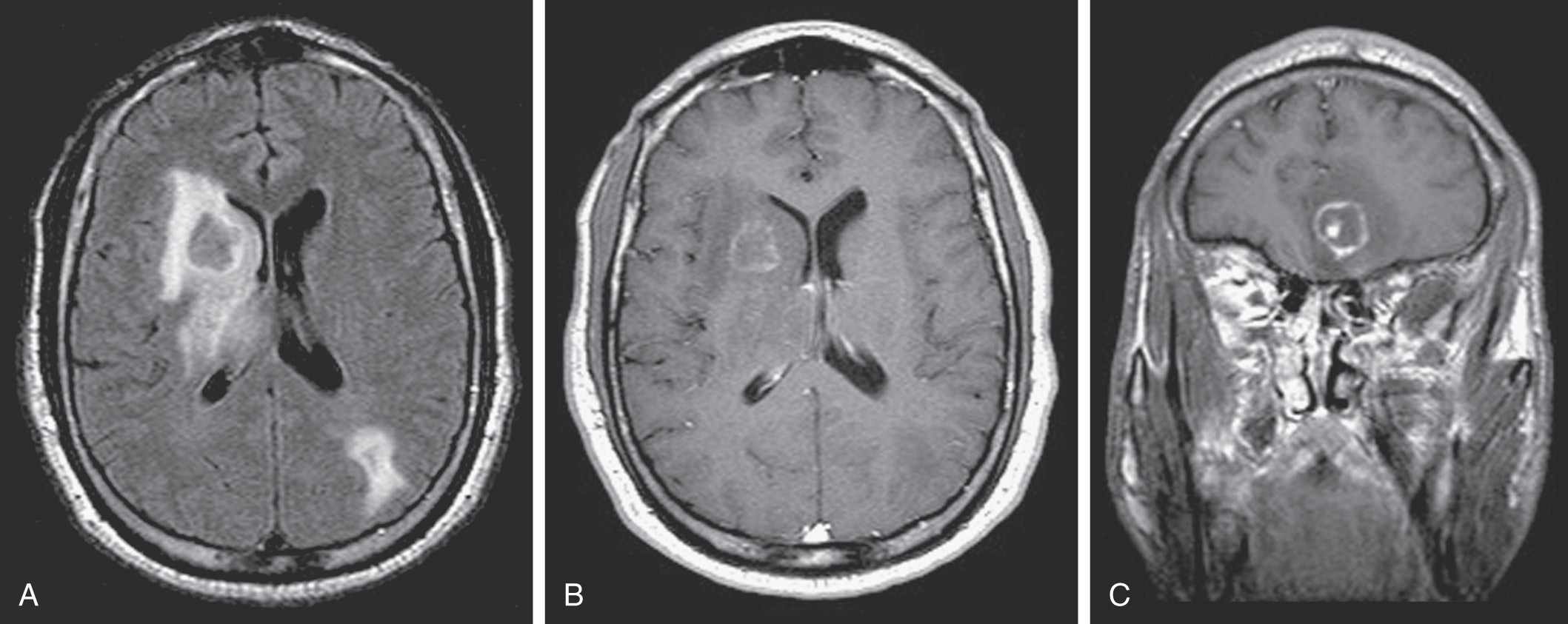
  • •MRI or CT of head for encephalopathy or focal CNS complications (e.g., toxoplasmosis [Fig. E2], lymphoma)
  • •Chest radiography or CT to aid in the diagnosis of Pneumocystis jiroveci pneumonia (PJP), TB, or bacterial pneumonia

Figure E2 Toxoplasmosis.

A, A fluid-attenuated inversion recovery image shows isointense lesions in the right basal ganglia and left parietal lobe with surrounding edema. B, A postgadolinium T1-weighted image shows faint rim enhancement of right basal ganglia lesion. C, A postgadolinium T1-weighted image shows a typical "target" sign in a left frontal lobe lesion.

(From Soto JA, Lucey BC: Emergency radiology: the requisites, ed 2, Philadelphia, 2017, Elsevier.)

Treatment ⬆ ⬇

The most important aspect in management of AIDS due to HIV infection is the timely initiation of antiretroviral therapy. Antiretroviral therapy should be initiated regardless of CD4 cell count.

Nonpharmacologic Therapy

  • •Maintain adequate caloric intake.
  • •Encourage good oral hygiene and regular dental care.
  • •Avoid high-risk behaviors that increase the risk of other potential pathogens-use condoms, avoid sharing needles, etc.
  • •Update vaccines-particularly Tdap (tetanus, diphtheria, and pertussis), pneumococcal, meningococcal, and hepatitis A/B vaccines along with annual influenza vaccines. COVID-19 vaccination is recommended.
  • •Avoid administration of any live attenuated vaccines that may be a risk to these immunocompromised patients (e.g., MMR, varicella). (See "Section V" for immunization schedules for HIV-infected children.)
  • •When feasible, avoid activities that might increase risk of exposure to opportunistic infections (e.g., cleaning out a cat litter box [toxoplasmosis], getting scratched by a cat [Bartonella infections], exposure to pet reptiles [salmonellosis], traveling to developing countries [cryptosporidiosis, tuberculosis], eating undercooked foods and drinking from unsafe water supplies, etc.). A description of enteropathogens causing infections in HIV-infected patients is provided in Table 3.

TABLE 3 Organisms That Cause Gastrointestinal Tract Infections in Patients With HIV/AIDS

Organisms
EsophagusCandida albicansa
Cytomegalovirusa
Herpes simplex virusa
HepatobiliaryCytomegalovirus
Cryptosporidiuma
Hepatotropic viruses
Mycobacterium avium complexa
Small intestineCampylobacter species
Cytomegalovirusa
Cryptosporidiuma
Giardia lambliaa
Isospora bellia
Mycobacterium avium complexa
Microsporidiaa (Enterocytozoon bieneusi and Encephalitozoon intestinalis)
Salmonella speciesa
Enteroaggregative E. coli
Strongyloides stercoralis
Large intestineCampylobacter species
Clostridium difficile
Cytomegalovirusa
Entamoeba histolytica
Herpes simplex virusa
Salmonella speciesa
Enteroaggregative E. coli
Shigella species

a Diseases of the gastrointestinal tract that fulfill the Centers for Disease Control and Prevention surveillance case definition of AIDS.

From Cherry JD et al: Feigin and Cherry’s pediatric infectious diseases, ed 8, Philadelphia, 2019, Elsevier.

Acute General Rx

Acute management of opportunistic infections is summarized in Table 4 and reviewed elsewhere in this text under specific AIDS-related disorders. For management of AIDS-related malignancies, please refer to the specific malignancy elsewhere in this text.

TABLE 4 Treatments for the Most Common Opportunistic Infections in Critically Ill HIV-Positive Patients

Opportunistic InfectionFirst-Line Regimen for Severe CasesMain AlternativesAdjunctive TherapiesTiming of cART Introduction
PCP pneumonia
  • TMP (15-20 mg/kg/d) and SMX (75-100 mg/kg/d) IV q6h or q8h
  • Switch to PO after clinical improvement
  • Total treatment duration: 3 wk (then switch to secondary prophylaxis dosinga)
  • No leucovorin supplementationb
Pentamidine 4 mg/kg IV once daily (patients with TMP or SMX adverse events such as allergy or hemolysis because of glucose-6-phosphate dehydrogenase deficiency)
  • Corticosteroids if PaO2<70 mm Hg (room air)
  • Prednisone PO:
  • Days 1-5: 40 mg BID
  • Days 6-10: 40 mg daily
  • Days 11-21: 20 mg daily
  • Alternative:
  • methylprednisolone IV (75% of prednisone dose)
Within 2 wk
Tuberculosis (drug-susceptible M. tuberculosis)
  • •Intensive phase (2 mo): isoniazid + rifampin or rifabutin + pyrazinamide + ethambutol
  • •Continuation phase: isoniazid + rifampin or rifabutin
  • •Total treatment duration:
  • Pulmonary TB: 6-9 mo
  • Extrapulmonary TB with CNS involvement: 9-12 mo
  • Extrapulmonary TB with bone or joint involvement: 6-9 mo
  • Extrapulmonary TB at other sites: 6 mo
Consult an ID specialist
  • •CNS disease: dexamethasone 0.3-0.4 mg/kg/d for 2-4 wk, then taper by 0.1 mg/kg per wk until 0.1 mg/kg, then 4 mg/d and taper by 1 mg/wk; total duration of 12 wk
  • •Pericardial disease: prednisone or prednisolone (e.g., 60 mg PO daily with weekly tapering over 6 wk)
  • •Active TB without CNS involvement:
  • •Within 2 wk for patients with CD4 cells <50/mL
  • •Within 8 wk for patients with CD4 cells ≥50/mL
  • •Active TB with CNS involvement: deferred initiation (high risk of severe IRIS)
  • •Close collaboration with ID specialists
Toxoplasmosis
  • •Pyrimethamine 200 mg PO once, then pyrimethamine 50-75 mg PO daily + sulfadiazine 1000-1500 mg PO q6h + leucovorin 10-25 mg PO daily
  • •Total treatment duration: at least 6 wk (then switch to chronic maintenance therapya)
  • •Pyrimethamine (with leucovorin) plus clindamycin 600 mg IV or PO q6h
  • •TMP 5 mg/kg and SMX 25 mg/kg IV or PO BID
  • •Corticosteroids if cerebral lesions with mass effect
  • •Anticonvulsants only if seizures (no primary prevention)
  • •No recommendation, because of insufficient data (usually within 2-3 wk)
Cryptococcus infections
  • •Induction therapy (at least 2 wk): liposomal amphotericin B 3-4 mg/kg IV daily plus flucytosine 25 mg/kg QID
  • •Consolidation therapy (at least 8 wk) after clinical improvement and sterilization of CSF cultures: fluconazole 400 mg PO or IV once daily
  • •Maintenance therapy (at least 1 yr): fluconazole 200 mg PO
Induction therapy: fluconazole (400-800 mg/d) may replace either liposomal amphotericin B or flucytosineCorticosteroids may be deleterious in patients with CNS disease and are not recommended.
  • CNS involvement: 2-10 wk after initiation of specific therapy (longer delay if high intracranial pressure)
  • Other localizations: 2-4 wk after initiation of specific therapy
Histoplasmosis
  • Induction therapy (at least 2 wk):
  • liposomal amphotericin B 3 mg/kg/d IV-If confirmed meningitis: increase dosage to 5 mg/kg/d and extend induction to 4-6 wk
  • Maintenance therapy: itraconazole 200 mg PO TID for 3 days, then BID for at least 12 mo
Fluconazole (400 md/d PO) may replace itraconazole for long-term suppressive therapy-As soon as possible
Disseminated MAC diseaseClarithromycin 500 mg PO twice daily + ethambutol 15 mg/kg PO daily or azithromycin 500-600 mg + ethambutol 15 mg/kg PO dailyConsult an infectious disease specialist-After completion of the second week of MAC-directed therapy
Addition of a third or fourth drug (rifabutin, amikacin, fluoroquinolone) should be considered for patients with CD41 T cells <50/mL, high mycobacterial loads (>2 log CFU/ml of blood), or in the absence of effective cART
CMV infection
  • Ganciclovir 5 mg/kg IV q12h
  • Role of oral valganciclovir: not established
  • Optimal treatment duration: not established
Foscarnet 60 mg/kg IV q8h or 90 mg/kg IV q12h-No recommendation, because of insufficient data (usually within 2 wk)
PMLcART--As soon as possible

BID, Twice a day; cART, Combination antiretroviral therapy; CFU, colony-forming unit; CMV, cytomegalovirus; CNS, central nervous system; CSF, cerebrospinal fluid; HIV, human immunodeficiency virus; IRIS, immune reconstitution inflammatory syndrome; IV, intravenous, intravenously; MAC, Mycobacterium avium complex; PaO2, arterial partial pressure of oxygen; PCP, Pneumocystis jiroveci; PML, progressive multifocal encephalopathy (caused by JC virus); PO, per os; SMX, sulfamethoxazole; TB, tuberculosis; TID, three times a day; TMP, trimethoprim. Society of America for the management of opportunistic infections in adults with HIV.

a Consider discontinuation in patients with CD4 cells >200/mL under cART.

b Leucovorin supplementation does not efficiently prevent myelosuppression and may be associated with treatment failure.

Adapted from the guidelines of the Centers for Disease Control and Prevention, the National Institutes of Health, and the Infectious Diseases Society. In Vincent JL et al: Textbook of critical care, ed 8, Philadelphia, 2024, Elsevier.

Chronic Rx

For all HIV-infected patients, particularly those meeting the case definition of AIDS:

  • •Preventive therapy for Pneumocystis jiroveci pneumonia, Mycobacterium avium complex (MAC) disease, and Toxoplasma gondii encephalitis are summarized in Table 5. With appropriate antiretroviral therapy, many patients experience substantial restoration of cellular immune function. Preventive therapy for Pneumocystis jiroveci can be safely stopped if the CD4 cell count rises above 200 for at least 3 mo.
  • •Based on the Department of Health and Human Services (DHHS) Guidelines, active antiretroviral therapy (ART) should be started regardless of CD4 cell count. Individuals with CD4 cell counts <350 and especially CD4 cell counts <200 should be strongly encouraged to start ART in a timely fashion.
  • •ART usually includes three-drug combinations of:
    1. 1.Nucleoside reverse transcriptase inhibitors (NRTI): Tenofovir disoproxil fumarate (TDF), tenofovir alafenamide fumarate (TAF), lamivudine (3TC), emtricitabine (FTC), and abacavir (ABC). Older drugs such as zidovudine (AZT), didanosine (ddI), and stavudine (d4T) are generally not recommended.
    2. 2.Protease inhibitors (PI): Darunavir (preferred) or atazanavir.
    3. 3.Nonnucleoside reverse transcriptase inhibitors (NNRTI): Nevirapine, efavirenz (EFV), etravirine, doravirine, and rilpivirine.
    4. 4.Integrase inhibitors: Raltegravir, elvitegravir, bictegravir, dolutegravir, and cabotegravir.
    5. 5.Others: Maraviroc, enfuvirtide, ibalizumab, and fostemsavir.

TABLE 5 Prophylaxis to Prevent First Episode of Selected Opportunistic Infections

PathogenIndicationFirst-Choice TherapyAlternative
Pneumocystis jiroveci pneumonia (PCP)
  • •CD4+<200 cells/mL or oropharyngeal candidiasis
  • •CD4+<14% or history of AIDS-defining illness
  • •CD4+>200 cells/mL but <250 cells/mL if monitoring is not possible every 1-3 mo
TMP-SMZ
  • TMP-SMZ
  • Dapsone
  • Dapsone + pyrimethamine + leucovorin
  • Aerosolized pentamidine
  • Atovaquone
  • Atovaquone + pyrimethamine + leucovorin
Toxoplasma gondii encephalitis
  • •Toxoplasma IgG-positive patients with CD4+ count <100 cells/mL
  • •Seronegative patients receiving PCP prophylaxis not active against toxoplasmosis should have Toxoplasma serology retested if CD4+ count declines to <100 cells/mL
  • •Initiate prophylaxis if seroconversion occurs.
TMP-SMZ
  • TMP-SMZ
  • Dapsone + pyrimethamine + leucovorin
  • Dapsone + pyrimethamine + leucovorin
  • Atovaquone ± pyrimethamine + leucovorin
Disseminated Mycobacterium avium complex (MAC) disease
  • •CD4+ count <50 cells/mL (after active MAC infection is ruled out)
Azithromycin or ClarithromycinRifabutin (adjust dose on basis of ART interactions); rule out active TB before rifabutin is started.

ART, Antiretroviral therapy; TB, tuberculosis; TMP-SMZ, trimethoprim-sulfamethoxazole.

Adapted from Kaplan JE et al: Guidelines for prevention and treatment of opportunistic infections in HIV-infected adults and adolescents: recommendations from CDC, the National Institutes of Health, and the HIV Medicine Association of the Infectious Diseases Society of America. Available at https://www.cdc.gov/mmwr/preview/mmwrhtml/rr5804a1.htm. In Walls RM et al: Rosen’s emergency medicine, concepts and clinical practice, ed 10, Philadelphia, 2023, Elsevier.

The medications ritonavir or cobicistat are usually used in combination with other protease inhibitors or integrase inhibitors to obtain more sustained drug levels. Usual initial dosing regimens include two NRTIs and an NNRTI or PI or integrase inhibitor. Two-drug regimens with dolutegravir and lamivudine can be considered in certain clinical situations. Currently, integrase inhibitors are recommended as first-line drugs because of tolerability. Examples of initial regimens recommended by the guidelines:

  • •Bictegravir/tenofovir alafenamide/emtricitabine
  • •Dolutegravir/abacavir/lamivudine (in patients who are HLA-B5701 NEGATIVE)
  • •Dolutegravir plus tenofovir/emtricitabine (tenofovir-based formulations can include tenofovir disoproxil fumarate [TDF] or tenofovir alafenamide [TAF])
  • •Dolutegravir and lamivudine (two-drug regimen). This regimen should not be used in individuals with HIV RNA >500,000 copies/ml, HBV coinfection, or in whom ART is to be started before the results of HIV genotypic resistance testing for reverse transcriptase or HBV testing are available.

All these drugs have unique and class-specific side effects and require careful and expert follow-up to achieve optimal antiviral effects, ensure compliance, and maintain efficacy. Antiviral response should be monitored by baseline HIV viral load and CD4 count and repeat measurement at 2 wk and 4 wk into treatment and then periodically (3 to 6 mo) to ensure viral suppression.

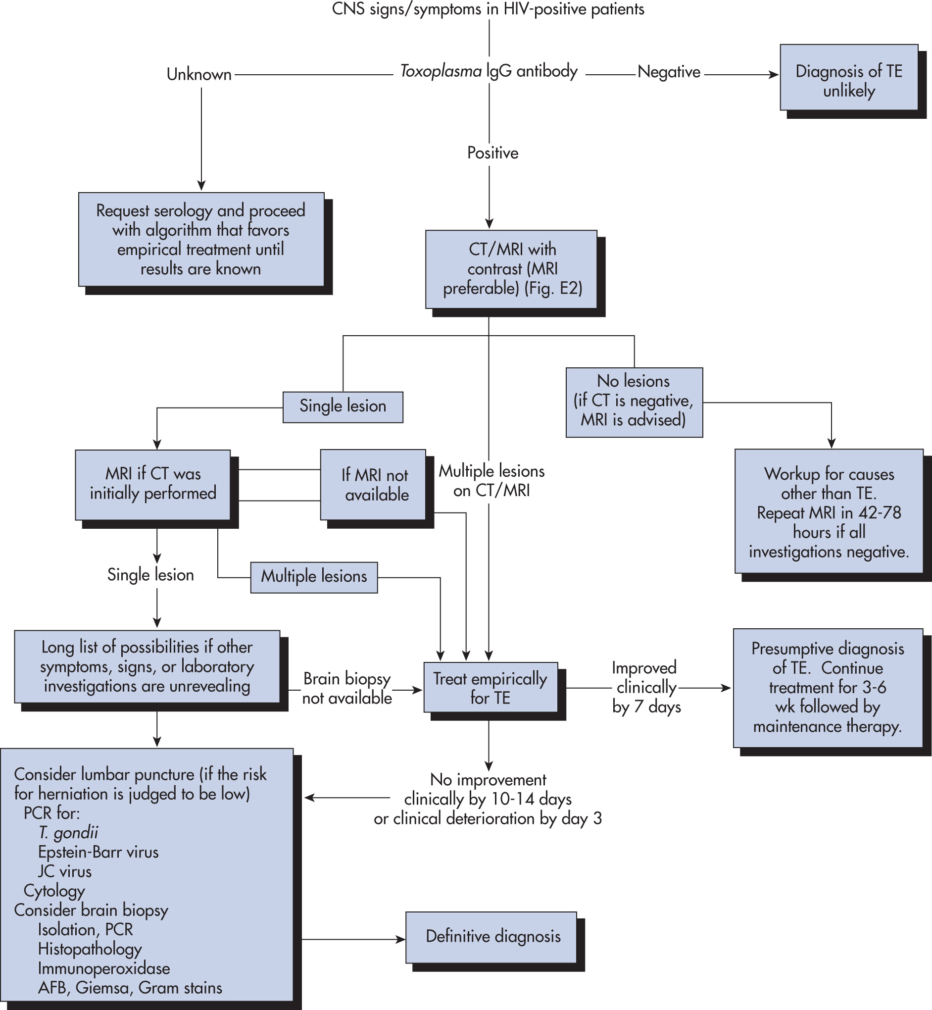
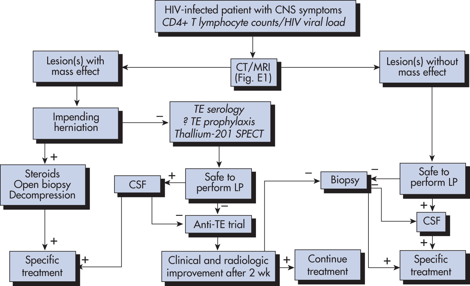
  • •The approach to a patient with CNS signs and symptoms is described in Fig. E3, and Fig. E4 describes the management of CNS mass lesions.
  • •Genotypic resistance testing is strongly encouraged for all patients initiating treatment and for any patient failing antiretroviral therapy. Poor adherence to therapy, however, often underlies virologic failure.

Figure E3 Diagnostic Approach and Management Algorithm for Human Immunodeficiency Virus (HIV)-Infected Patients with Central Nervous System (CNS) Symptoms or Signs that Might Potentially Be Toxoplasmic Encephalitis (TE)

!!flowchart!!

AFB, Acid-fast bacilli; CT, computed tomography; IgG, immunoglobulin G; MRI, magnetic resonance imaging; PCR, polymerase chain reaction.

(From Bennett JE et al: Mandell, Douglas, and Bennett’s principles and practice of infectious diseases, ed 9, Philadelphia, 2020, Elsevier.)

Figure E4 Management of the Human Immunodeficiency Virus (HIV) Type 1-Infected Patient with Central Nervous System (CNS) Mass Lesions
!!flowchart!!

The elements in italics represent data that contribute to the decision-making process (see text for details). CSF, Cerebrospinal fluid; CT, computed tomography; LP, lumbar puncture; MRI, magnetic resonance imaging; SPECT, single-photon emission computed tomography; TE, Toxoplasma encephalitis.

(From Bennett JE et al: Mandell, Douglas, and Bennett’s principles and practice of infectious diseases, ed 9, Philadelphia, 2020, Elsevier.)

Disposition

The outlook for HIV has changed radically since the advent of ART from an essentially fatal disease to a chronic medical illness compatible with long-term survival and remarkably good quality of life. Patients should be aggressively treated for severe illnesses as outcomes following ICU admissions remain good. This is accomplished through expert and continuous follow-up, use of ART, and careful detail to compliance to medications and lifestyle modification.

Referral

All patients with HIV should be referred to a physician knowledgeable and experienced in the management of the disease and its complications.

Pearls & Considerations ⬆

Related Content